Alexa R Heaton, Nathaniel J Burkard, Paul M Sondel, Melissa C Skala
{"title":"Quantifying <i>in vivo</i> collagen reorganization during immunotherapy in murine melanoma with second harmonic generation imaging.","authors":"Alexa R Heaton, Nathaniel J Burkard, Paul M Sondel, Melissa C Skala","doi":"10.1117/1.bios.1.1.015004","DOIUrl":null,"url":null,"abstract":"<p><strong>Significance: </strong>Increased collagen linearization and deposition during tumorigenesis can impede immune cell infiltration and lead to tumor metastasis. Although melanoma is well studied in immunotherapy research, studies that quantify collagen changes during melanoma progression and treatment are lacking.</p><p><strong>Aim: </strong>We aim to image <i>in vivo</i> collagen in preclinical melanoma models during immunotherapy and quantify the collagen phenotype in treated and control mice.</p><p><strong>Approach: </strong>Second-harmonic generation imaging of collagen was performed in mouse melanoma tumors <i>in vivo</i> over a treatment time course. Animals were treated with a curative radiation and immunotherapy combination. Collagen morphology was quantified over time at an image and single-fiber level using CurveAlign and CT-FIRE software.</p><p><strong>Results: </strong>In immunotherapy-treated mice, collagen was reorganized toward a healthy phenotype, including shorter, wider, curlier collagen fibers, with modestly higher collagen density. Temporally, collagen fiber straightness and length changed late in treatment (days 9 and 12), while width and density changed early (day 6) compared with control mice. Single-fiber collagen features calculated in CT-FIRE were the most sensitive to the changes among treatment groups compared with bulk collagen features.</p><p><strong>Conclusions: </strong>Quantitative second-harmonic generation imaging can provide insight into collagen dynamics <i>in vivo</i> during immunotherapy, with key implications in improving immunotherapy response in melanoma and other cancers.</p>","PeriodicalId":519981,"journal":{"name":"Biophotonics discovery","volume":"1 1","pages":""},"PeriodicalIF":0.0000,"publicationDate":"2024-05-01","publicationTypes":"Journal Article","fieldsOfStudy":null,"isOpenAccess":false,"openAccessPdf":"https://www.ncbi.nlm.nih.gov/pmc/articles/PMC11247620/pdf/","citationCount":"0","resultStr":null,"platform":"Semanticscholar","paperid":null,"PeriodicalName":"Biophotonics discovery","FirstCategoryId":"1085","ListUrlMain":"https://doi.org/10.1117/1.bios.1.1.015004","RegionNum":0,"RegionCategory":null,"ArticlePicture":[],"TitleCN":null,"AbstractTextCN":null,"PMCID":null,"EPubDate":"2024/5/20 0:00:00","PubModel":"Epub","JCR":"","JCRName":"","Score":null,"Total":0}
引用次数: 0
Abstract
Significance: Increased collagen linearization and deposition during tumorigenesis can impede immune cell infiltration and lead to tumor metastasis. Although melanoma is well studied in immunotherapy research, studies that quantify collagen changes during melanoma progression and treatment are lacking.
Aim: We aim to image in vivo collagen in preclinical melanoma models during immunotherapy and quantify the collagen phenotype in treated and control mice.
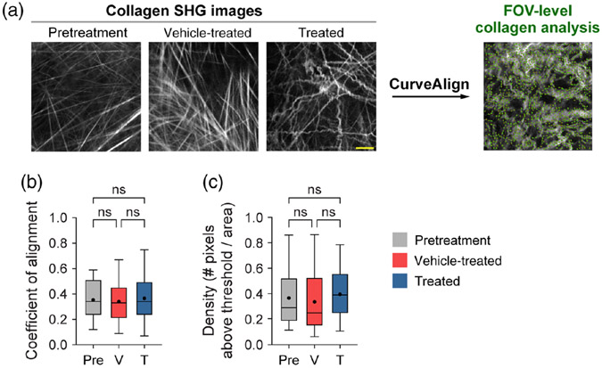
Approach: Second-harmonic generation imaging of collagen was performed in mouse melanoma tumors in vivo over a treatment time course. Animals were treated with a curative radiation and immunotherapy combination. Collagen morphology was quantified over time at an image and single-fiber level using CurveAlign and CT-FIRE software.
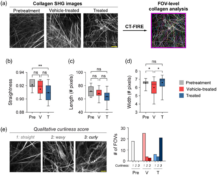
Results: In immunotherapy-treated mice, collagen was reorganized toward a healthy phenotype, including shorter, wider, curlier collagen fibers, with modestly higher collagen density. Temporally, collagen fiber straightness and length changed late in treatment (days 9 and 12), while width and density changed early (day 6) compared with control mice. Single-fiber collagen features calculated in CT-FIRE were the most sensitive to the changes among treatment groups compared with bulk collagen features.
Conclusions: Quantitative second-harmonic generation imaging can provide insight into collagen dynamics in vivo during immunotherapy, with key implications in improving immunotherapy response in melanoma and other cancers.


 分享
分享

 求助内容:
求助内容: 应助结果提醒方式:
 扫码关注我们
