Brianae Garcia, Anis Hasnaoui, Prakash V A K Ramdass
{"title":"Lupus Erythematosus Tumidus Misdiagnosed as Erythema Nodosum from Coccidioidomycosis.","authors":"Brianae Garcia, Anis Hasnaoui, Prakash V A K Ramdass","doi":"10.1159/000538737","DOIUrl":null,"url":null,"abstract":"<p><strong>Introduction: </strong>Lupus erythematosus tumidus (LET) is a rare photosensitive dermatosis that is categorized as intermittent cutaneous lupus erythematosus. It shares clinical similarities and histopathological features with other skin disorders, such as erythema nodosum, lymphocytic infiltrate of Jessner, and reticular erythematous mucinosis, thus making diagnosis quite challenging. We present a patient with LET whose diagnosis was confirmed after seeing several doctors.</p><p><strong>Case presentation: </strong>A 52-year-old Hispanic female presented with tender erythematous nodules on her thighs for approximately 1 month. She was suspected of having erythema nodosum secondary to coccidioidomycosis and was prescribed fluconazole 200 mg for 30 days but showed no improvement. However, histopathological and direct immunofluorescence tests later confirmed a diagnosis of LET. The patient was treated with hydroxychloroquine, and the lesions improved remarkably after 2 weeks.</p><p><strong>Conclusion: </strong>LET is a rare dermatosis that closely resembles other dermatologic conditions such as erythema nodosum, lymphocytic infiltrate of Jessner, and reticular erythematous mucinosis. Diagnosis based on clinical features alone should be avoided, and ideally, treatment should only be initiated after confirmatory histopathological testing.</p>","PeriodicalId":9619,"journal":{"name":"Case Reports in Dermatology","volume":"16 1","pages":"128-132"},"PeriodicalIF":0.7000,"publicationDate":"2024-05-23","publicationTypes":"Journal Article","fieldsOfStudy":null,"isOpenAccess":false,"openAccessPdf":"https://www.ncbi.nlm.nih.gov/pmc/articles/PMC11250662/pdf/","citationCount":"0","resultStr":null,"platform":"Semanticscholar","paperid":null,"PeriodicalName":"Case Reports in Dermatology","FirstCategoryId":"1085","ListUrlMain":"https://doi.org/10.1159/000538737","RegionNum":0,"RegionCategory":null,"ArticlePicture":[],"TitleCN":null,"AbstractTextCN":null,"PMCID":null,"EPubDate":"2024/1/1 0:00:00","PubModel":"eCollection","JCR":"Q4","JCRName":"DERMATOLOGY","Score":null,"Total":0}
引用次数: 0
Abstract
Introduction: Lupus erythematosus tumidus (LET) is a rare photosensitive dermatosis that is categorized as intermittent cutaneous lupus erythematosus. It shares clinical similarities and histopathological features with other skin disorders, such as erythema nodosum, lymphocytic infiltrate of Jessner, and reticular erythematous mucinosis, thus making diagnosis quite challenging. We present a patient with LET whose diagnosis was confirmed after seeing several doctors.
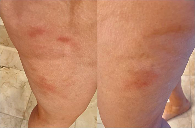
Case presentation: A 52-year-old Hispanic female presented with tender erythematous nodules on her thighs for approximately 1 month. She was suspected of having erythema nodosum secondary to coccidioidomycosis and was prescribed fluconazole 200 mg for 30 days but showed no improvement. However, histopathological and direct immunofluorescence tests later confirmed a diagnosis of LET. The patient was treated with hydroxychloroquine, and the lesions improved remarkably after 2 weeks.
Conclusion: LET is a rare dermatosis that closely resembles other dermatologic conditions such as erythema nodosum, lymphocytic infiltrate of Jessner, and reticular erythematous mucinosis. Diagnosis based on clinical features alone should be avoided, and ideally, treatment should only be initiated after confirmatory histopathological testing.


 分享
分享
 求助内容:
求助内容: 应助结果提醒方式:
 扫码关注我们
