Cedar A Slovacek, Yang Li, Maria Hurley, Ramona Beshad, Michael L Bernstein
{"title":"Pilomatrix Carcinoma: A Benign-Mimic with Malignant Consequences - A Case Report and Review of the Current Literature.","authors":"Cedar A Slovacek, Yang Li, Maria Hurley, Ramona Beshad, Michael L Bernstein","doi":"10.1159/000539123","DOIUrl":null,"url":null,"abstract":"<p><strong>Introduction: </strong>Pilomatrix carcinomas (PMXCs) are uncommon, locally aggressive tumors with high recurrence rates, metastatic potential, and fewer than 130 cases reported in the literature. Typically, they present as an unassuming, firm, dermal swelling and therefore are frequently mistaken for more common, benign masses, leading to undertreatment which can cause local invasion and metastatic spread. Diagnosis relies on excision with pathologic analysis; however once diagnosed, there are no current recommendations to guide treatment or surveillance for recurrence or metastases.</p><p><strong>Case presentation: </strong>Here, we present a case of one of these rare tumors. Our case describes a 1.5 × 2.5 cm firm, mobile mass at the supraorbital rim in an otherwise healthy, young patient. Prior to removal, we suspected a benign pathology; however, excision proved difficult and pathologic diagnosis was consistent with PMXC. Following discussion with tumor board, decision was made to perform Mohs micrographic surgery and staging via CT scans with regular follow-up and surveillance scans.</p><p><strong>Conclusion: </strong>PMXCs are exceedingly rare diagnoses and present like many benign lesions. Therefore, we elected to document this case to encourage providers to keep these biologically aggressive tumors on their list of differential diagnoses in an unsuspecting mass, as well as to provide our own recommendations for treatment and screening for recurrence and metastatic spread.</p>","PeriodicalId":9619,"journal":{"name":"Case Reports in Dermatology","volume":"16 1","pages":"156-163"},"PeriodicalIF":0.8000,"publicationDate":"2024-06-18","publicationTypes":"Journal Article","fieldsOfStudy":null,"isOpenAccess":false,"openAccessPdf":"https://www.ncbi.nlm.nih.gov/pmc/articles/PMC11250398/pdf/","citationCount":"0","resultStr":null,"platform":"Semanticscholar","paperid":null,"PeriodicalName":"Case Reports in Dermatology","FirstCategoryId":"1085","ListUrlMain":"https://doi.org/10.1159/000539123","RegionNum":0,"RegionCategory":null,"ArticlePicture":[],"TitleCN":null,"AbstractTextCN":null,"PMCID":null,"EPubDate":"2024/1/1 0:00:00","PubModel":"eCollection","JCR":"Q4","JCRName":"DERMATOLOGY","Score":null,"Total":0}
引用次数: 0
Abstract
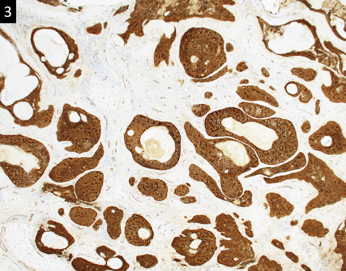
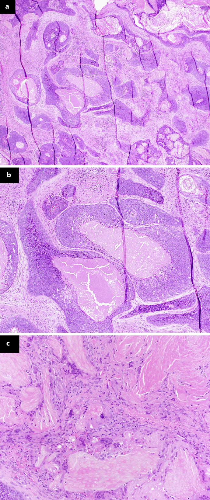
Introduction: Pilomatrix carcinomas (PMXCs) are uncommon, locally aggressive tumors with high recurrence rates, metastatic potential, and fewer than 130 cases reported in the literature. Typically, they present as an unassuming, firm, dermal swelling and therefore are frequently mistaken for more common, benign masses, leading to undertreatment which can cause local invasion and metastatic spread. Diagnosis relies on excision with pathologic analysis; however once diagnosed, there are no current recommendations to guide treatment or surveillance for recurrence or metastases.
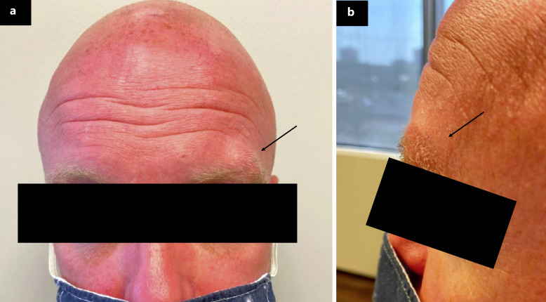
Case presentation: Here, we present a case of one of these rare tumors. Our case describes a 1.5 × 2.5 cm firm, mobile mass at the supraorbital rim in an otherwise healthy, young patient. Prior to removal, we suspected a benign pathology; however, excision proved difficult and pathologic diagnosis was consistent with PMXC. Following discussion with tumor board, decision was made to perform Mohs micrographic surgery and staging via CT scans with regular follow-up and surveillance scans.
Conclusion: PMXCs are exceedingly rare diagnoses and present like many benign lesions. Therefore, we elected to document this case to encourage providers to keep these biologically aggressive tumors on their list of differential diagnoses in an unsuspecting mass, as well as to provide our own recommendations for treatment and screening for recurrence and metastatic spread.


 分享
分享
 求助内容:
求助内容: 应助结果提醒方式:
 扫码关注我们
