Satheesha B Nayak, Surekha Devadasa Shetty, Ashwini Aithal P
{"title":"Vesico-obturator venous plexus - a rare termination of obturator vein.","authors":"Satheesha B Nayak, Surekha Devadasa Shetty, Ashwini Aithal P","doi":"10.1007/s00276-024-03434-6","DOIUrl":null,"url":null,"abstract":"<p><p>Obturator vein usually terminates into the internal iliac vein. Its variations are the cause major bleeding problems in pelvic surgeries. We observed a rare variation in the termination of the right obturator vein. There was a duplication of right obturator vein. Both obturator veins entered the pelvic cavity through the obturator foramen and joined with two vesical veins to form a vesico-obturator plexus. This plexus surrounded the internal iliac artery and terminated into the internal iliac vein. Awareness of this rare variation could be of importance to anatomists, radiologists, gynaecologists, urologists, and orthopaedic surgeons. The plexus might lead to hazardous bleeding in pelvic lymph node clearance procedures, hernia surgeries, gynaecological and orthopaedic procedures in this region. The pelvic fractures too can provoke dramatic retroperitoneal hematomas related to these veins injuries.</p>","PeriodicalId":49461,"journal":{"name":"Surgical and Radiologic Anatomy","volume":" ","pages":"1491-1493"},"PeriodicalIF":1.2000,"publicationDate":"2024-09-01","publicationTypes":"Journal Article","fieldsOfStudy":null,"isOpenAccess":false,"openAccessPdf":"","citationCount":"0","resultStr":null,"platform":"Semanticscholar","paperid":null,"PeriodicalName":"Surgical and Radiologic Anatomy","FirstCategoryId":"3","ListUrlMain":"https://doi.org/10.1007/s00276-024-03434-6","RegionNum":4,"RegionCategory":"医学","ArticlePicture":[],"TitleCN":null,"AbstractTextCN":null,"PMCID":null,"EPubDate":"2024/7/17 0:00:00","PubModel":"Epub","JCR":"Q2","JCRName":"Medicine","Score":null,"Total":0}
引用次数: 0
Abstract
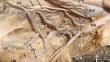
Obturator vein usually terminates into the internal iliac vein. Its variations are the cause major bleeding problems in pelvic surgeries. We observed a rare variation in the termination of the right obturator vein. There was a duplication of right obturator vein. Both obturator veins entered the pelvic cavity through the obturator foramen and joined with two vesical veins to form a vesico-obturator plexus. This plexus surrounded the internal iliac artery and terminated into the internal iliac vein. Awareness of this rare variation could be of importance to anatomists, radiologists, gynaecologists, urologists, and orthopaedic surgeons. The plexus might lead to hazardous bleeding in pelvic lymph node clearance procedures, hernia surgeries, gynaecological and orthopaedic procedures in this region. The pelvic fractures too can provoke dramatic retroperitoneal hematomas related to these veins injuries.
期刊介绍:
Anatomy is a morphological science which cannot fail to interest the clinician. The practical application of anatomical research to clinical problems necessitates special adaptation and selectivity in choosing from numerous international works. Although there is a tendency to believe that meaningful advances in anatomy are unlikely, constant revision is necessary. Surgical and Radiologic Anatomy, the first international journal of Clinical anatomy has been created in this spirit.
Its goal is to serve clinicians, regardless of speciality-physicians, surgeons, radiologists or other specialists-as an indispensable aid with which they can improve their knowledge of anatomy. Each issue includes: Original papers, review articles, articles on the anatomical bases of medical, surgical and radiological techniques, articles of normal radiologic anatomy, brief reviews of anatomical publications of clinical interest.
Particular attention is given to high quality illustrations, which are indispensable for a better understanding of anatomical problems.
Surgical and Radiologic Anatomy is a journal written by anatomists for clinicians with a special interest in anatomy.


 分享
分享
 求助内容:
求助内容: 应助结果提醒方式:
 扫码关注我们
