Chujie Zhang, Juncong Ma, Chang Liu, Xianliang Yan
{"title":"The protective effect of karanjin against sepsis-induced acute lung injury in mice is involved in the suppression of the TLR4 pathway","authors":"Chujie Zhang, Juncong Ma, Chang Liu, Xianliang Yan","doi":"10.1111/cbdd.14579","DOIUrl":null,"url":null,"abstract":"<p>Sepsis-induced acute lung injury (ALI) is a severe complication of sepsis. Karanjin, a natural flavonoid compound, has been proved to have anti-inflammatory function, but its role in sepsis-stimulated ALI is uncertain. Herein, the effect of karanjin on sepsis-stimulated ALI was investigated. We built a mouse model of lipopolysaccharide (LPS)-stimulated ALI. The histopathological morphology of lung tissues was scrutinized by hematoxylin–eosin (H&E) staining. The lung injury score and lung wet/dry weight ratio were detected. The myeloperoxidase (MPO) activity and malondialdehyde (MDA) content were scrutinized by commercial kits. Murine alveolar lung epithelial (MLE-12) cells were treated with LPS to mimic a cellular model of ALI. The cell viability was scrutinized by the CCK-8 assay. The contents of proinflammatory cytokines were scrutinized by qRT-PCR and ELISA. The TLR4 and MyD88 contents were scrutinized by qRT-PCR and western blotting. Results showed that karanjin alleviated LPS-stimulated ALI in mice by inhibiting lung tissue lesions, edema, and oxidative stress. Moreover, karanjin inhibited LPS-stimulated inflammation and TLR4 pathway activation in mice. However, treatment with GSK1795091, an agonist of TLR4, attenuated the effects of karanjin on LPS-induced ALI. Furthermore, karanjin repressed LPS-stimulated inflammatory response and TLR4 pathway activation in MLE-12 cells. Overexpression of TLR4 attenuated karanjin effects on LPS-stimulated inflammatory responses in MLE-12 cells. In conclusion, karanjin repressed sepsis-stimulated ALI in mice by suppressing the TLR4 pathway.</p>","PeriodicalId":143,"journal":{"name":"Chemical Biology & Drug Design","volume":"104 1","pages":""},"PeriodicalIF":3.2000,"publicationDate":"2024-07-16","publicationTypes":"Journal Article","fieldsOfStudy":null,"isOpenAccess":false,"openAccessPdf":"","citationCount":"0","resultStr":null,"platform":"Semanticscholar","paperid":null,"PeriodicalName":"Chemical Biology & Drug Design","FirstCategoryId":"3","ListUrlMain":"https://onlinelibrary.wiley.com/doi/10.1111/cbdd.14579","RegionNum":4,"RegionCategory":"医学","ArticlePicture":[],"TitleCN":null,"AbstractTextCN":null,"PMCID":null,"EPubDate":"","PubModel":"","JCR":"Q2","JCRName":"BIOCHEMISTRY & MOLECULAR BIOLOGY","Score":null,"Total":0}
引用次数: 0
Abstract
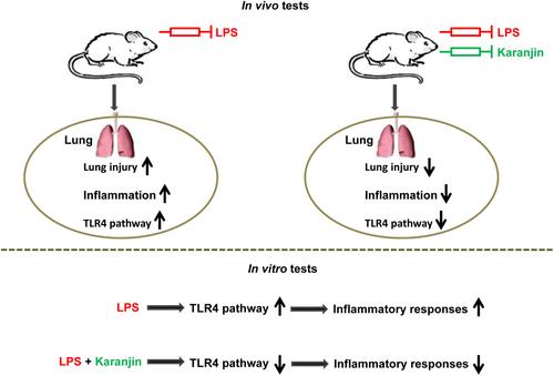
Sepsis-induced acute lung injury (ALI) is a severe complication of sepsis. Karanjin, a natural flavonoid compound, has been proved to have anti-inflammatory function, but its role in sepsis-stimulated ALI is uncertain. Herein, the effect of karanjin on sepsis-stimulated ALI was investigated. We built a mouse model of lipopolysaccharide (LPS)-stimulated ALI. The histopathological morphology of lung tissues was scrutinized by hematoxylin–eosin (H&E) staining. The lung injury score and lung wet/dry weight ratio were detected. The myeloperoxidase (MPO) activity and malondialdehyde (MDA) content were scrutinized by commercial kits. Murine alveolar lung epithelial (MLE-12) cells were treated with LPS to mimic a cellular model of ALI. The cell viability was scrutinized by the CCK-8 assay. The contents of proinflammatory cytokines were scrutinized by qRT-PCR and ELISA. The TLR4 and MyD88 contents were scrutinized by qRT-PCR and western blotting. Results showed that karanjin alleviated LPS-stimulated ALI in mice by inhibiting lung tissue lesions, edema, and oxidative stress. Moreover, karanjin inhibited LPS-stimulated inflammation and TLR4 pathway activation in mice. However, treatment with GSK1795091, an agonist of TLR4, attenuated the effects of karanjin on LPS-induced ALI. Furthermore, karanjin repressed LPS-stimulated inflammatory response and TLR4 pathway activation in MLE-12 cells. Overexpression of TLR4 attenuated karanjin effects on LPS-stimulated inflammatory responses in MLE-12 cells. In conclusion, karanjin repressed sepsis-stimulated ALI in mice by suppressing the TLR4 pathway.
期刊介绍:
Chemical Biology & Drug Design is a peer-reviewed scientific journal that is dedicated to the advancement of innovative science, technology and medicine with a focus on the multidisciplinary fields of chemical biology and drug design. It is the aim of Chemical Biology & Drug Design to capture significant research and drug discovery that highlights new concepts, insight and new findings within the scope of chemical biology and drug design.


 分享
分享
 求助内容:
求助内容: 应助结果提醒方式:
 扫码关注我们
