{"title":"Structural insights into the assembly pathway of the Helicobacter pylori CagT4SS outer membrane core complex","authors":"","doi":"10.1016/j.str.2024.06.019","DOIUrl":null,"url":null,"abstract":"<p>Cag type IV secretion system (CagT4SS) translocates oncoprotein cytotoxin-associated gene A (CagA) into host cells and plays a key role in the pathogenesis of <em>Helicobacter pylori</em>. The structure of the outer membrane core complex (OMCC) in CagT4SS consists of CagX, CagY, CagM, CagT, and Cag3 in a stoichiometric ratio of 1:1:2:2:5 with 14-fold symmetry. However, the assembly pathway of OMCC remains elusive. Here, we report the crystal structures of CagT and Cag3-CagT complex, and the structural dynamics of Cag3 and CagT using hydrogen deuterium exchange-mass spectrometry (HDX-MS). The interwoven interaction of Cag3 and CagT involves conformational changes of CagT and β strand swapping. In conjunction with biochemical and biophysical assays, we further demonstrate the different oligomerization states of Cag3 and Cag3-CagT complex. Additionally, the association with CagM requires the pre-formation of Cag3-CagT complex. These results demonstrate the generation of different intermediate sub-assemblies and their structural flexibility, potentially representing different building blocks for OMCC assembly.</p>","PeriodicalId":22168,"journal":{"name":"Structure","volume":"7 1","pages":""},"PeriodicalIF":4.3000,"publicationDate":"2024-07-19","publicationTypes":"Journal Article","fieldsOfStudy":null,"isOpenAccess":false,"openAccessPdf":"","citationCount":"0","resultStr":null,"platform":"Semanticscholar","paperid":null,"PeriodicalName":"Structure","FirstCategoryId":"99","ListUrlMain":"https://doi.org/10.1016/j.str.2024.06.019","RegionNum":2,"RegionCategory":"生物学","ArticlePicture":[],"TitleCN":null,"AbstractTextCN":null,"PMCID":null,"EPubDate":"","PubModel":"","JCR":"Q2","JCRName":"BIOCHEMISTRY & MOLECULAR BIOLOGY","Score":null,"Total":0}
引用次数: 0
Abstract
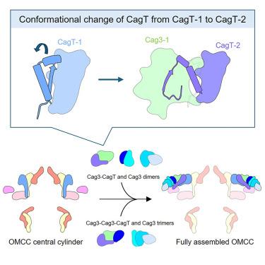
Cag type IV secretion system (CagT4SS) translocates oncoprotein cytotoxin-associated gene A (CagA) into host cells and plays a key role in the pathogenesis of Helicobacter pylori. The structure of the outer membrane core complex (OMCC) in CagT4SS consists of CagX, CagY, CagM, CagT, and Cag3 in a stoichiometric ratio of 1:1:2:2:5 with 14-fold symmetry. However, the assembly pathway of OMCC remains elusive. Here, we report the crystal structures of CagT and Cag3-CagT complex, and the structural dynamics of Cag3 and CagT using hydrogen deuterium exchange-mass spectrometry (HDX-MS). The interwoven interaction of Cag3 and CagT involves conformational changes of CagT and β strand swapping. In conjunction with biochemical and biophysical assays, we further demonstrate the different oligomerization states of Cag3 and Cag3-CagT complex. Additionally, the association with CagM requires the pre-formation of Cag3-CagT complex. These results demonstrate the generation of different intermediate sub-assemblies and their structural flexibility, potentially representing different building blocks for OMCC assembly.
期刊介绍:
Structure aims to publish papers of exceptional interest in the field of structural biology. The journal strives to be essential reading for structural biologists, as well as biologists and biochemists that are interested in macromolecular structure and function. Structure strongly encourages the submission of manuscripts that present structural and molecular insights into biological function and mechanism. Other reports that address fundamental questions in structural biology, such as structure-based examinations of protein evolution, folding, and/or design, will also be considered. We will consider the application of any method, experimental or computational, at high or low resolution, to conduct structural investigations, as long as the method is appropriate for the biological, functional, and mechanistic question(s) being addressed. Likewise, reports describing single-molecule analysis of biological mechanisms are welcome.
In general, the editors encourage submission of experimental structural studies that are enriched by an analysis of structure-activity relationships and will not consider studies that solely report structural information unless the structure or analysis is of exceptional and broad interest. Studies reporting only homology models, de novo models, or molecular dynamics simulations are also discouraged unless the models are informed by or validated by novel experimental data; rationalization of a large body of existing experimental evidence and making testable predictions based on a model or simulation is often not considered sufficient.


 分享
分享
 求助内容:
求助内容: 应助结果提醒方式:
 扫码关注我们
