Andaleeb A Ahmed, Robina Matyal, Feroze Mahmood, Ruby Feng, Graham B Berry, Scott Gilleland, Kamal R Khabbaz
{"title":"Impact of left ventricular outflow tract flow acceleration on aortic valve area calculation in patients with aortic stenosis","authors":"Andaleeb A Ahmed, Robina Matyal, Feroze Mahmood, Ruby Feng, Graham B Berry, Scott Gilleland, Kamal R Khabbaz","doi":"","DOIUrl":null,"url":null,"abstract":"<p><strong>Objective: </strong>Due to its circular shape, the area of the proximal left ventricular tract (PLVOT) adjacent to aortic valve can be derived from a single linear diameter. This is also the location of flow acceleration (FA) during systole, and pulse wave Doppler (PWD) sample volume in the PLVOT can lead to overestimation of velocity (V<sub>1</sub>) and the aortic valve area (AVA). Therefore, it is recommended to derive V<sub>1</sub> from a region of laminar flow in the elliptical shaped distal LVOT (away from the annulus). Besides being inconsistent with the assumptions of continuity equation (CE), spatial difference in the location of flow and area measurement can result in inaccurate AVA calculation. We evaluated the impact of FA in the PLVOT on the accuracy of AVA by continuity equation (CE) in patients with aortic stenosis (AS).</p><p><strong>Methods: </strong>CE-based AVA calculations were performed in patients with AS once with PWD-derived velocity time integral (VTI) in the distal LVOT (VTI<sub>LVOT</sub>) and then in the PLVOT to obtain a FA velocity profile (FA-VTI<sub>LVOT</sub>) for each patient. A paired sample t-test (P < 0.05) was conducted to compare the impact of FA-VTI<sub>LVOT</sub> and VTI<sub>LVOT</sub> on the calculation of AVA.</p><p><strong>Result: </strong>There were 46 patients in the study. There was a 30.3% increase in the peak FA-VTI<sub>LVOT</sub> as compared to the peak VTI<sub>LVOT</sub> and AVA obtained by FA-VTI<sub>LVOT</sub> was 29.1% higher than obtained by VTI<sub>LVOT</sub>.</p><p><strong>Conclusion: </strong>Accuracy of AVA can be significantly impacted by FA in the PLVOT. LVOT area should be measured with 3D imaging in the distal LVOT.</p>","PeriodicalId":45749,"journal":{"name":"Echo Research and Practice","volume":"6 4","pages":"97-103"},"PeriodicalIF":2.4000,"publicationDate":"2019-11-04","publicationTypes":"Journal Article","fieldsOfStudy":null,"isOpenAccess":false,"openAccessPdf":"https://www.ncbi.nlm.nih.gov/pmc/articles/PMC6826166/pdf/","citationCount":"0","resultStr":null,"platform":"Semanticscholar","paperid":null,"PeriodicalName":"Echo Research and Practice","FirstCategoryId":"1085","ListUrlMain":"","RegionNum":0,"RegionCategory":null,"ArticlePicture":[],"TitleCN":null,"AbstractTextCN":null,"PMCID":null,"EPubDate":"","PubModel":"","JCR":"Q2","JCRName":"CARDIAC & CARDIOVASCULAR SYSTEMS","Score":null,"Total":0}
引用次数: 0
Abstract
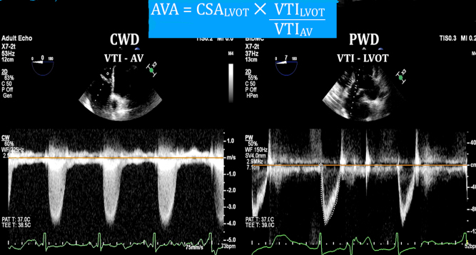
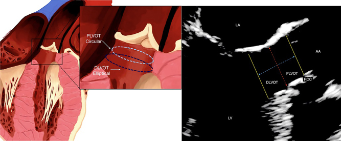
Objective: Due to its circular shape, the area of the proximal left ventricular tract (PLVOT) adjacent to aortic valve can be derived from a single linear diameter. This is also the location of flow acceleration (FA) during systole, and pulse wave Doppler (PWD) sample volume in the PLVOT can lead to overestimation of velocity (V1) and the aortic valve area (AVA). Therefore, it is recommended to derive V1 from a region of laminar flow in the elliptical shaped distal LVOT (away from the annulus). Besides being inconsistent with the assumptions of continuity equation (CE), spatial difference in the location of flow and area measurement can result in inaccurate AVA calculation. We evaluated the impact of FA in the PLVOT on the accuracy of AVA by continuity equation (CE) in patients with aortic stenosis (AS).
Methods: CE-based AVA calculations were performed in patients with AS once with PWD-derived velocity time integral (VTI) in the distal LVOT (VTILVOT) and then in the PLVOT to obtain a FA velocity profile (FA-VTILVOT) for each patient. A paired sample t-test (P < 0.05) was conducted to compare the impact of FA-VTILVOT and VTILVOT on the calculation of AVA.
Result: There were 46 patients in the study. There was a 30.3% increase in the peak FA-VTILVOT as compared to the peak VTILVOT and AVA obtained by FA-VTILVOT was 29.1% higher than obtained by VTILVOT.
Conclusion: Accuracy of AVA can be significantly impacted by FA in the PLVOT. LVOT area should be measured with 3D imaging in the distal LVOT.
目的:由于左心室近端通道(PLVOT)呈圆形,其邻近主动脉瓣的面积可通过单一线性直径得出。这也是收缩期血流加速(FA)的位置,PLVOT 中的脉搏波多普勒(PWD)样本量会导致速度(V1)和主动脉瓣面积(AVA)被高估。因此,建议从椭圆形 LVOT 远端(远离瓣环)的层流区域推导 V1。除了与连续性方程(CE)的假设不一致外,血流位置和面积测量的空间差异也会导致 AVA 计算不准确。我们评估了 PLVOT 中的 FA 对主动脉瓣狭窄(AS)患者用连续性方程(CE)计算 AVA 的准确性的影响:方法:我们对 AS 患者进行了基于 CE 的 AVA 计算,计算时先在 LVOT 远端(VTILVOT)使用 PWD 导出的速度时间积分(VTI),然后在 PLVOT 获取 FA 速度曲线(FA-VTILVOT)。对 AVA 的计算结果进行配对样本 t 检验(P LVOT 和 VTILVOT):共有 46 名患者参与了研究。与 VTILVOT 峰值相比,FA-VTILVOT 峰值增加了 30.3%,FA-VTILVOT 获得的 AVA 比 VTILVOT 高 29.1%:结论:PLVOT 中的 FA 会严重影响 AVA 的准确性。结论:PLVOT 的 FA 会严重影响 AVA 的准确性。
期刊介绍:
Echo Research and Practice aims to be the premier international journal for physicians, sonographers, nurses and other allied health professionals practising echocardiography and other cardiac imaging modalities. This open-access journal publishes quality clinical and basic research, reviews, videos, education materials and selected high-interest case reports and videos across all echocardiography modalities and disciplines, including paediatrics, anaesthetics, general practice, acute medicine and intensive care. Multi-modality studies primarily featuring the use of cardiac ultrasound in clinical practice, in association with Cardiac Computed Tomography, Cardiovascular Magnetic Resonance or Nuclear Cardiology are of interest. Topics include, but are not limited to: 2D echocardiography 3D echocardiography Comparative imaging techniques – CCT, CMR and Nuclear Cardiology Congenital heart disease, including foetal echocardiography Contrast echocardiography Critical care echocardiography Deformation imaging Doppler echocardiography Interventional echocardiography Intracardiac echocardiography Intraoperative echocardiography Prosthetic valves Stress echocardiography Technical innovations Transoesophageal echocardiography Valve disease.

 分享
分享


 求助内容:
求助内容: 应助结果提醒方式:
 扫码关注我们
