{"title":"Investigation of choroidal structure changes after intravitreal anti-VEGF therapy for retinal vein occlusion.","authors":"Erdem Dursun, Baki Derhem, Seval Çobanoğlu, Tevfik Oğurel","doi":"10.1007/s00417-024-06562-2","DOIUrl":null,"url":null,"abstract":"<p><strong>Background: </strong>We aimed to investigate the effect of retinal vein occlusion (RVO) on the posterior segment structures of the eye and its changes with intravitreal anti-Vascular Endothelial Growth Factor (VEGF) treatment.</p><p><strong>Methods: </strong>This prospective longitudinal study included 29 eyes of 29 patients with RVO (17 males and 12 females) followed for 6 months. The best corrected visual acuity (BCVA), macula, choroid ticknesses and choroidal vascularity index (CVI) obtained by spectral-domain optical coherence tomography were recorded at baseline and the first, third, and sixth months after the first injection. Results were compared with fellow eyes (non-affected eyes) and age- and sex-matched controls.</p><p><strong>Results: </strong>BCVA increased significantly in the 6th month, more in the first month of injection (p < 0.05 for each). Central macular tickness, subfoveal choroid tickness, stromal and total area of choroid decreased significantly after injection (p < 0.05 for each). CVI values increased significantly, especially in the 1st month after injection (p < 0.05 for each). In eyes with Branch RVO, there was a significant decrease in the macular thickness of the occlusive areas with treatment, while there was no statistically significant change in the non-occlusive macular thickness.</p><p><strong>Conclusion: </strong>Observation of changes in choroidal structure may be useful to assess the activity of RVO and predict the efficacy of anti-VEGF therapy.</p>","PeriodicalId":12795,"journal":{"name":"Graefe’s Archive for Clinical and Experimental Ophthalmology","volume":" ","pages":"3837-3845"},"PeriodicalIF":2.4000,"publicationDate":"2024-12-01","publicationTypes":"Journal Article","fieldsOfStudy":null,"isOpenAccess":false,"openAccessPdf":"https://www.ncbi.nlm.nih.gov/pmc/articles/PMC11608387/pdf/","citationCount":"0","resultStr":null,"platform":"Semanticscholar","paperid":null,"PeriodicalName":"Graefe’s Archive for Clinical and Experimental Ophthalmology","FirstCategoryId":"3","ListUrlMain":"https://doi.org/10.1007/s00417-024-06562-2","RegionNum":3,"RegionCategory":"医学","ArticlePicture":[],"TitleCN":null,"AbstractTextCN":null,"PMCID":null,"EPubDate":"2024/7/22 0:00:00","PubModel":"Epub","JCR":"Q2","JCRName":"OPHTHALMOLOGY","Score":null,"Total":0}
引用次数: 0
Abstract
Background: We aimed to investigate the effect of retinal vein occlusion (RVO) on the posterior segment structures of the eye and its changes with intravitreal anti-Vascular Endothelial Growth Factor (VEGF) treatment.
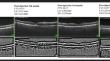
Methods: This prospective longitudinal study included 29 eyes of 29 patients with RVO (17 males and 12 females) followed for 6 months. The best corrected visual acuity (BCVA), macula, choroid ticknesses and choroidal vascularity index (CVI) obtained by spectral-domain optical coherence tomography were recorded at baseline and the first, third, and sixth months after the first injection. Results were compared with fellow eyes (non-affected eyes) and age- and sex-matched controls.
Results: BCVA increased significantly in the 6th month, more in the first month of injection (p < 0.05 for each). Central macular tickness, subfoveal choroid tickness, stromal and total area of choroid decreased significantly after injection (p < 0.05 for each). CVI values increased significantly, especially in the 1st month after injection (p < 0.05 for each). In eyes with Branch RVO, there was a significant decrease in the macular thickness of the occlusive areas with treatment, while there was no statistically significant change in the non-occlusive macular thickness.
Conclusion: Observation of changes in choroidal structure may be useful to assess the activity of RVO and predict the efficacy of anti-VEGF therapy.
期刊介绍:
Graefe''s Archive for Clinical and Experimental Ophthalmology is a distinguished international journal that presents original clinical reports and clini-cally relevant experimental studies. Founded in 1854 by Albrecht von Graefe to serve as a source of useful clinical information and a stimulus for discussion, the journal has published articles by leading ophthalmologists and vision research scientists for more than a century. With peer review by an international Editorial Board and prompt English-language publication, Graefe''s Archive provides rapid dissemination of clinical and clinically related experimental information.


 分享
分享
 求助内容:
求助内容: 应助结果提醒方式:
 扫码关注我们
