{"title":"Investigation of abdominal artery delineation by photon-counting detector CT.","authors":"Takashi Ota, Hiromitsu Onishi, Toshihide Itoh, Hideyuki Fukui, Takahiro Tsuboyama, Atsushi Nakamoto, Yukihiro Enchi, Mitsuaki Tatsumi, Noriyuki Tomiyama","doi":"10.1007/s11547-024-01858-z","DOIUrl":null,"url":null,"abstract":"<p><strong>Objectives: </strong>To evaluate the ability of 50-keV virtual monoenergetic images (VMI) to depict abdominal arteries in abdominal CT angiography (CTA) compared with 70-keV VMI with photon-counting detector CT (PCD-CT).</p><p><strong>Methods: </strong>Fifty consecutive patients who underwent multiphase abdominal scans between March and April 2023 were included. Signal-to-noise ratio (SNR) and contrast-to-noise ratio (CNR) were quantitatively assessed for the abdominal aorta (AA), celiac artery (CeA), superior mesenteric artery (SMA), renal artery (RA), and right hepatic artery (RHA) at both 50- and 70-keV VMI. In addition, 3D images from CTA were analyzed to measure arterial lengths and evaluate the visualization of distal branches.</p><p><strong>Results: </strong>Significantly higher SNR and CNR were observed at 50-keV compared to 70-keV VMI for all arteries: AA (36.54 and 48.28 vs. 25.70 and 28.46), CeA (22.39 and 48.38 vs. 19.09 and 29.15), SMA (23.34 and 49.34 vs. 19.67 and 29.71), RA (22.88 and 48.84 vs. 20.15 and 29.41), and RHA (14.38 and 44.41 vs. 13.45 and 27.18), all p < 0.05. Arterial lengths were also significantly longer at 50-keV: RHA (192.6 vs. 180.3 mm), SMA (230.9 vs. 216.5 mm), and RA (95.9 vs. 92.0 mm), all p < 0.001.</p><p><strong>Conclusion: </strong>In abdominal CTA with PCD-CT, 50-keV VMI demonstrated superior quantitative image quality compared to 70-keV VMI. In addition, 50-keV VMI 3D CTA allowed better visualization of abdominal artery branches, highlighting its potential clinical advantage for improved imaging and detailed assessment of abdominal arteries.</p>","PeriodicalId":20817,"journal":{"name":"Radiologia Medica","volume":" ","pages":"1265-1274"},"PeriodicalIF":4.8000,"publicationDate":"2024-09-01","publicationTypes":"Journal Article","fieldsOfStudy":null,"isOpenAccess":false,"openAccessPdf":"https://www.ncbi.nlm.nih.gov/pmc/articles/PMC11379784/pdf/","citationCount":"0","resultStr":null,"platform":"Semanticscholar","paperid":null,"PeriodicalName":"Radiologia Medica","FirstCategoryId":"3","ListUrlMain":"https://doi.org/10.1007/s11547-024-01858-z","RegionNum":1,"RegionCategory":"医学","ArticlePicture":[],"TitleCN":null,"AbstractTextCN":null,"PMCID":null,"EPubDate":"2024/7/24 0:00:00","PubModel":"Epub","JCR":"Q1","JCRName":"RADIOLOGY, NUCLEAR MEDICINE & MEDICAL IMAGING","Score":null,"Total":0}
引用次数: 0
Abstract
Objectives: To evaluate the ability of 50-keV virtual monoenergetic images (VMI) to depict abdominal arteries in abdominal CT angiography (CTA) compared with 70-keV VMI with photon-counting detector CT (PCD-CT).
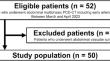
Methods: Fifty consecutive patients who underwent multiphase abdominal scans between March and April 2023 were included. Signal-to-noise ratio (SNR) and contrast-to-noise ratio (CNR) were quantitatively assessed for the abdominal aorta (AA), celiac artery (CeA), superior mesenteric artery (SMA), renal artery (RA), and right hepatic artery (RHA) at both 50- and 70-keV VMI. In addition, 3D images from CTA were analyzed to measure arterial lengths and evaluate the visualization of distal branches.
Results: Significantly higher SNR and CNR were observed at 50-keV compared to 70-keV VMI for all arteries: AA (36.54 and 48.28 vs. 25.70 and 28.46), CeA (22.39 and 48.38 vs. 19.09 and 29.15), SMA (23.34 and 49.34 vs. 19.67 and 29.71), RA (22.88 and 48.84 vs. 20.15 and 29.41), and RHA (14.38 and 44.41 vs. 13.45 and 27.18), all p < 0.05. Arterial lengths were also significantly longer at 50-keV: RHA (192.6 vs. 180.3 mm), SMA (230.9 vs. 216.5 mm), and RA (95.9 vs. 92.0 mm), all p < 0.001.
Conclusion: In abdominal CTA with PCD-CT, 50-keV VMI demonstrated superior quantitative image quality compared to 70-keV VMI. In addition, 50-keV VMI 3D CTA allowed better visualization of abdominal artery branches, highlighting its potential clinical advantage for improved imaging and detailed assessment of abdominal arteries.
期刊介绍:
Felice Perussia founded La radiologia medica in 1914. It is a peer-reviewed journal and serves as the official journal of the Italian Society of Medical and Interventional Radiology (SIRM). The primary purpose of the journal is to disseminate information related to Radiology, especially advancements in diagnostic imaging and related disciplines. La radiologia medica welcomes original research on both fundamental and clinical aspects of modern radiology, with a particular focus on diagnostic and interventional imaging techniques. It also covers topics such as radiotherapy, nuclear medicine, radiobiology, health physics, and artificial intelligence in the context of clinical implications. The journal includes various types of contributions such as original articles, review articles, editorials, short reports, and letters to the editor. With an esteemed Editorial Board and a selection of insightful reports, the journal is an indispensable resource for radiologists and professionals in related fields. Ultimately, La radiologia medica aims to serve as a platform for international collaboration and knowledge sharing within the radiological community.


 分享
分享
 求助内容:
求助内容: 应助结果提醒方式:
 扫码关注我们
