Claudin18.2-Targeted SPECT/CT Imaging for Gastric Cancer: Preclinical Evaluation and Clinical Translation of the 99mTc-Labeled Nanobody (PHG102) Radiotracer
{"title":"Claudin18.2-Targeted SPECT/CT Imaging for Gastric Cancer: Preclinical Evaluation and Clinical Translation of the 99mTc-Labeled Nanobody (PHG102) Radiotracer","authors":"Zhidong Bai, Xin Xie, Chenzhen Li, Yuchen Wang, Yuanbo Wang, Huijie Li, Rui Gao, Bing Jia","doi":"10.1021/acsptsci.4c00280","DOIUrl":null,"url":null,"abstract":"Claudin18.2 (CLDN18.2) has emerged as a significant target in the treatment of advanced gastric cancer. The screening of patients positive for CLDN18.2 is crucial for the effective application of targeted therapies specific to CLND18.2. In this study, we developed a novel nanobody-based probe, [<sup>99m</sup>Tc]Tc-PHG102, for use in nuclear medicine. We analyzed its radiochemical yield and stability to ensure accurate probe characterization. Additionally, we assessed the probe’s affinity and specificity toward the CLDN18.2 target and evaluated its efficacy in the BGC823<sup>18.2</sup> xenograft model for SPECT/CT imaging of gastric cancer. The binding of [<sup>99m</sup>Tc]Tc-PHG102 to HEK-293T<sup>18.2</sup> and BGC823<sup>18.2</sup> cells was notably higher than its binding to HEK-293T<sup>18.1</sup>, HEK-293T, and BGC823 cells, with bound values of 12.87 ± 1.46%, 6.16 ± 0.34%, 1.25 ± 0.22%, 1.14 ± 0.26%, and 1.32 ± 0.07% AD, respectively. The binding ability of [<sup>99m</sup>Tc]Tc-PHG102 was significantly different between CLDN18.2-positive and negative cells (<i>P</i> < 0.001). Imaging results demonstrated a time-dependent tumor accumulation of the radiotracer. Notably, at 0.5 h postinjection, rapid accumulation was observed with an average tumor uptake of 4.63 ± 0.81% ID/cc (<i>n</i> = 3), resulting in clear tumor visualization. By 1 h postinjection, as [<sup>99m</sup>Tc]Tc-PHG102 was rapidly metabolized, a decrease in uptake by other organs was noted. Preliminary clinical imaging trials further confirmed the safety and effectiveness of the probe, indicating specificity for lesions expressing CLDN18.2 in gastric cancer and favorable in vivo metabolic properties. In conclusion, the nanobody-based probe [<sup>99m</sup>Tc]Tc-PHG102 proves to be a safe and effective tool for detecting CLDN18.2 expression levels in gastric cancer tumors and for screening CLDN18.2-positive patients.","PeriodicalId":501473,"journal":{"name":"ACS Pharmacology & Translational Science","volume":"43 1","pages":""},"PeriodicalIF":0.0000,"publicationDate":"2024-07-20","publicationTypes":"Journal Article","fieldsOfStudy":null,"isOpenAccess":false,"openAccessPdf":"","citationCount":"0","resultStr":null,"platform":"Semanticscholar","paperid":null,"PeriodicalName":"ACS Pharmacology & Translational Science","FirstCategoryId":"1085","ListUrlMain":"https://doi.org/10.1021/acsptsci.4c00280","RegionNum":0,"RegionCategory":null,"ArticlePicture":[],"TitleCN":null,"AbstractTextCN":null,"PMCID":null,"EPubDate":"","PubModel":"","JCR":"","JCRName":"","Score":null,"Total":0}
引用次数: 0
Abstract
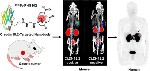
Claudin18.2 (CLDN18.2) has emerged as a significant target in the treatment of advanced gastric cancer. The screening of patients positive for CLDN18.2 is crucial for the effective application of targeted therapies specific to CLND18.2. In this study, we developed a novel nanobody-based probe, [99mTc]Tc-PHG102, for use in nuclear medicine. We analyzed its radiochemical yield and stability to ensure accurate probe characterization. Additionally, we assessed the probe’s affinity and specificity toward the CLDN18.2 target and evaluated its efficacy in the BGC82318.2 xenograft model for SPECT/CT imaging of gastric cancer. The binding of [99mTc]Tc-PHG102 to HEK-293T18.2 and BGC82318.2 cells was notably higher than its binding to HEK-293T18.1, HEK-293T, and BGC823 cells, with bound values of 12.87 ± 1.46%, 6.16 ± 0.34%, 1.25 ± 0.22%, 1.14 ± 0.26%, and 1.32 ± 0.07% AD, respectively. The binding ability of [99mTc]Tc-PHG102 was significantly different between CLDN18.2-positive and negative cells (P < 0.001). Imaging results demonstrated a time-dependent tumor accumulation of the radiotracer. Notably, at 0.5 h postinjection, rapid accumulation was observed with an average tumor uptake of 4.63 ± 0.81% ID/cc (n = 3), resulting in clear tumor visualization. By 1 h postinjection, as [99mTc]Tc-PHG102 was rapidly metabolized, a decrease in uptake by other organs was noted. Preliminary clinical imaging trials further confirmed the safety and effectiveness of the probe, indicating specificity for lesions expressing CLDN18.2 in gastric cancer and favorable in vivo metabolic properties. In conclusion, the nanobody-based probe [99mTc]Tc-PHG102 proves to be a safe and effective tool for detecting CLDN18.2 expression levels in gastric cancer tumors and for screening CLDN18.2-positive patients.


 分享
分享

 求助内容:
求助内容: 应助结果提醒方式:
 扫码关注我们
