Haihai Jiang, Cheng Lin, Jingyi Chang, Xiaofang Zou, Jin Zhang, Jian Li
{"title":"Crystal structures of the 3C proteases from Coxsackievirus B3 and B4","authors":"Haihai Jiang, Cheng Lin, Jingyi Chang, Xiaofang Zou, Jin Zhang, Jian Li","doi":"10.1107/S2053230X24006915","DOIUrl":null,"url":null,"abstract":"<p>Enteroviruses cause a wide range of disorders with varying presentations and severities, and some enteroviruses have emerged as serious public health concerns. These include Coxsackievirus B3 (CVB3), an active causative agent of viral myocarditis, and Coxsackievirus B4 (CVB4), which may accelerate the progression of type 1 diabetes. The 3C proteases from CVB3 and CVB4 play important roles in the propagation of these viruses. In this study, the 3C proteases from CVB3 and CVB4 were expressed in <i>Escherichia coli</i> and purified by affinity chromatography and gel-filtration chromatography. The crystals of the CVB3 and CVB4 3C proteases diffracted to 2.10 and 2.01 Å resolution, respectively. The crystal structures were solved by the molecular-replacement method and contained a typical chymotrypsin-like fold and a conserved His40–Glu71–Cys147 catalytic triad. Comparison with the structures of 3C proteases from other enteroviruses revealed high similarity with minor differences, which will guide the design of 3C-targeting inhibitors with broad-spectrum properties.</p>","PeriodicalId":7029,"journal":{"name":"Acta crystallographica. Section F, Structural biology communications","volume":"80 8","pages":"183-190"},"PeriodicalIF":1.1000,"publicationDate":"2024-07-25","publicationTypes":"Journal Article","fieldsOfStudy":null,"isOpenAccess":false,"openAccessPdf":"","citationCount":"0","resultStr":null,"platform":"Semanticscholar","paperid":null,"PeriodicalName":"Acta crystallographica. Section F, Structural biology communications","FirstCategoryId":"99","ListUrlMain":"https://onlinelibrary.wiley.com/doi/10.1107/S2053230X24006915","RegionNum":4,"RegionCategory":"生物学","ArticlePicture":[],"TitleCN":null,"AbstractTextCN":null,"PMCID":null,"EPubDate":"","PubModel":"","JCR":"Q4","JCRName":"BIOCHEMICAL RESEARCH METHODS","Score":null,"Total":0}
引用次数: 0
Abstract
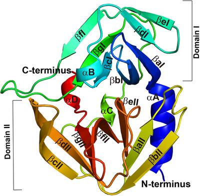
Enteroviruses cause a wide range of disorders with varying presentations and severities, and some enteroviruses have emerged as serious public health concerns. These include Coxsackievirus B3 (CVB3), an active causative agent of viral myocarditis, and Coxsackievirus B4 (CVB4), which may accelerate the progression of type 1 diabetes. The 3C proteases from CVB3 and CVB4 play important roles in the propagation of these viruses. In this study, the 3C proteases from CVB3 and CVB4 were expressed in Escherichia coli and purified by affinity chromatography and gel-filtration chromatography. The crystals of the CVB3 and CVB4 3C proteases diffracted to 2.10 and 2.01 Å resolution, respectively. The crystal structures were solved by the molecular-replacement method and contained a typical chymotrypsin-like fold and a conserved His40–Glu71–Cys147 catalytic triad. Comparison with the structures of 3C proteases from other enteroviruses revealed high similarity with minor differences, which will guide the design of 3C-targeting inhibitors with broad-spectrum properties.
期刊介绍:
Acta Crystallographica Section F is a rapid structural biology communications journal.
Articles on any aspect of structural biology, including structures determined using high-throughput methods or from iterative studies such as those used in the pharmaceutical industry, are welcomed by the journal.
The journal offers the option of open access, and all communications benefit from unlimited free use of colour illustrations and no page charges. Authors are encouraged to submit multimedia content for publication with their articles.
Acta Cryst. F has a dedicated online tool called publBio that is designed to make the preparation and submission of articles easier for authors.


 分享
分享
 求助内容:
求助内容: 应助结果提醒方式:
 扫码关注我们
