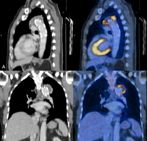
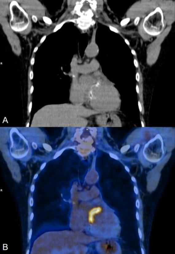
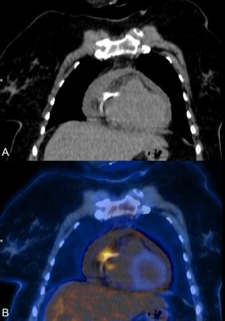
{"title":"<sup>18</sup>F-FDG PET CT in cardiac device infections - A Case series.","authors":"Awiral Saxena, Manjit Sarma, Bhagirath Bhad, Ramkesh Ratheesan, Padma Subramanyam, P Shanmuga Sundaram","doi":"10.22038/AOJNMB.2024.77504.1547","DOIUrl":null,"url":null,"abstract":"<p><p>With the increasing number of interventional cardiology procedures, the number of cardiac device infections (including pacemakers, prosthetic valves, coronary and aortic stents) have also increased. These infections can cause significant morbidity and can even lead to mortality if not managed promptly. If suspected clinically the first-line imaging modality is Trans-Thoracic Echocardiography, while Transesophageal Echocardiography is also used in selected cases. The confirmation of a cardiac device infection is mostly done with the help of blood or pus culture. Even though Echocardiography is a very efficient technique for the evaluation of the heart, it cannot differentiate infection from thrombus or fibrosis. With the increasing availability of Positron Emission Tomography CT (PET CT) machines worldwide, the use of <sup>18</sup>F-FDG PET CT for infection imaging has gained traction, especially for cardiac device infection. Most of the recent studies show a good diagnostic accuracy of <sup>18</sup>F-FDG PET CT with many of the recent diagnostic and management guidelines now acknowledging its role, especially in equivocal cases. We present six such cases where <sup>18</sup>F-FDG PET CT provided valuable information either for diagnosis, confirming the presence of infection, delineating extent, therapy response or sometimes even helping appropriate treatment decision making in patients with suspected cardiac device infection.</p>","PeriodicalId":8503,"journal":{"name":"Asia Oceania Journal of Nuclear Medicine and Biology","volume":"12 2","pages":"161-169"},"PeriodicalIF":0.0000,"publicationDate":"2024-01-01","publicationTypes":"Journal Article","fieldsOfStudy":null,"isOpenAccess":false,"openAccessPdf":"https://www.ncbi.nlm.nih.gov/pmc/articles/PMC11263774/pdf/","citationCount":"0","resultStr":null,"platform":"Semanticscholar","paperid":null,"PeriodicalName":"Asia Oceania Journal of Nuclear Medicine and Biology","FirstCategoryId":"1085","ListUrlMain":"https://doi.org/10.22038/AOJNMB.2024.77504.1547","RegionNum":0,"RegionCategory":null,"ArticlePicture":[],"TitleCN":null,"AbstractTextCN":null,"PMCID":null,"EPubDate":"","PubModel":"","JCR":"Q3","JCRName":"Medicine","Score":null,"Total":0}
引用次数: 0
Abstract
With the increasing number of interventional cardiology procedures, the number of cardiac device infections (including pacemakers, prosthetic valves, coronary and aortic stents) have also increased. These infections can cause significant morbidity and can even lead to mortality if not managed promptly. If suspected clinically the first-line imaging modality is Trans-Thoracic Echocardiography, while Transesophageal Echocardiography is also used in selected cases. The confirmation of a cardiac device infection is mostly done with the help of blood or pus culture. Even though Echocardiography is a very efficient technique for the evaluation of the heart, it cannot differentiate infection from thrombus or fibrosis. With the increasing availability of Positron Emission Tomography CT (PET CT) machines worldwide, the use of 18F-FDG PET CT for infection imaging has gained traction, especially for cardiac device infection. Most of the recent studies show a good diagnostic accuracy of 18F-FDG PET CT with many of the recent diagnostic and management guidelines now acknowledging its role, especially in equivocal cases. We present six such cases where 18F-FDG PET CT provided valuable information either for diagnosis, confirming the presence of infection, delineating extent, therapy response or sometimes even helping appropriate treatment decision making in patients with suspected cardiac device infection.
随着介入心脏病学手术数量的增加,心脏设备感染(包括起搏器、人工瓣膜、冠状动脉和主动脉支架)的数量也在增加。这些感染会导致严重的发病率,如果处理不及时甚至会导致死亡。如果临床上怀疑有心脏装置感染,第一线影像检查方法是经胸超声心动图,经食道超声心动图也可用于特定病例。心脏设备感染的确诊大多需要借助血液或脓液培养。尽管超声心动图是一种非常有效的心脏评估技术,但它无法区分感染与血栓或纤维化。随着正电子发射计算机断层扫描(PET CT)机在全球范围内的普及,18F-FDG PET CT 在感染成像中的应用越来越受到重视,尤其是在心脏设备感染方面。最近的大多数研究表明,18F-FDG PET CT 具有良好的诊断准确性,许多最新的诊断和管理指南都承认了它的作用,尤其是在诊断不明确的病例中。我们介绍了六个这样的病例,在这些病例中,18F-FDG PET CT 为疑似心脏设备感染患者的诊断、确诊感染的存在、划定感染范围、治疗反应,有时甚至帮助做出适当的治疗决策提供了有价值的信息。


 分享
分享
 求助内容:
求助内容: 应助结果提醒方式:
 扫码关注我们
