Mahdieh Parvizi, Mehrshad Abbasi, Hojjat Ahmadzadehfar, Abbas Tafakhori, Maryam Naseri, Ali Khalaj, Saeed Farzanehfar
{"title":"Bio-distribution study of Tc-99m HMPAO labeled platelet in healthy volunteer.","authors":"Mahdieh Parvizi, Mehrshad Abbasi, Hojjat Ahmadzadehfar, Abbas Tafakhori, Maryam Naseri, Ali Khalaj, Saeed Farzanehfar","doi":"10.22038/AOJNMB.2024.71620.1525","DOIUrl":null,"url":null,"abstract":"<p><strong>Objectives: </strong>The bio-distribution of Tc-99m HMPAO labeled platelets (LP), which could be used to image subtle thrombosis, is not reported in a human yet, which is the subject of the current study.</p><p><strong>Method: </strong>The platelets were extracted from 49 ml whole blood and labeled with Tc-99m HMPAO, then re-injected to the healthy volunteer. Anterior and posterior whole body imaging was done by a dual-head gamma camera 3, 18, 33, 46, 81, 124, 190 min and 15 hours after injection. Also a whole-body SPECT was done at 137 min post-injection. The area under the curves of the spleen, liver, left kidney, bladder, right lung, brain, and abdominal aorta ROIs was calculated to estimate the accumulation of labeled platelets within the organs.</p><p><strong>Results: </strong>The spleen was the target organ. The kidneys, liver, and heart were also remarkably visualized. The thyroid, stomach, bladder, or gastrointestinal (GI) uptake/activity was not significant. The stomach visualization was enhanced after ingestion at 60 min. The sagittal and lateral sinuses were delineated, and the background of the brain was very low. During the study, the area under the curve of activity was 738, 308, 302, 196, 230, 121, 79, 216, 529, 369, 162, and 54 counts. min/pixel for spleen, liver, heart, right lung, left kidney, right iliac artery, sagittal sinus, thyroid, bladder, stomach, GI, and background, respectively.</p><p><strong>Conclusion: </strong>The quality of the scan with low dose Tc-99m HMPAO LPs is optimal. We documented the bio-distribution of LPs. The optimal imaging time was 80-120 min post-injection when the free Tc-99m and GI transit were negligible. The sagittal and lateral sinuses were visualized enabling detection of possible clots in the vessels.</p>","PeriodicalId":8503,"journal":{"name":"Asia Oceania Journal of Nuclear Medicine and Biology","volume":"12 2","pages":"142-148"},"PeriodicalIF":0.0000,"publicationDate":"2024-01-01","publicationTypes":"Journal Article","fieldsOfStudy":null,"isOpenAccess":false,"openAccessPdf":"https://www.ncbi.nlm.nih.gov/pmc/articles/PMC11263780/pdf/","citationCount":"0","resultStr":null,"platform":"Semanticscholar","paperid":null,"PeriodicalName":"Asia Oceania Journal of Nuclear Medicine and Biology","FirstCategoryId":"1085","ListUrlMain":"https://doi.org/10.22038/AOJNMB.2024.71620.1525","RegionNum":0,"RegionCategory":null,"ArticlePicture":[],"TitleCN":null,"AbstractTextCN":null,"PMCID":null,"EPubDate":"","PubModel":"","JCR":"Q3","JCRName":"Medicine","Score":null,"Total":0}
引用次数: 0
Abstract
Objectives: The bio-distribution of Tc-99m HMPAO labeled platelets (LP), which could be used to image subtle thrombosis, is not reported in a human yet, which is the subject of the current study.
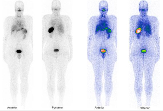
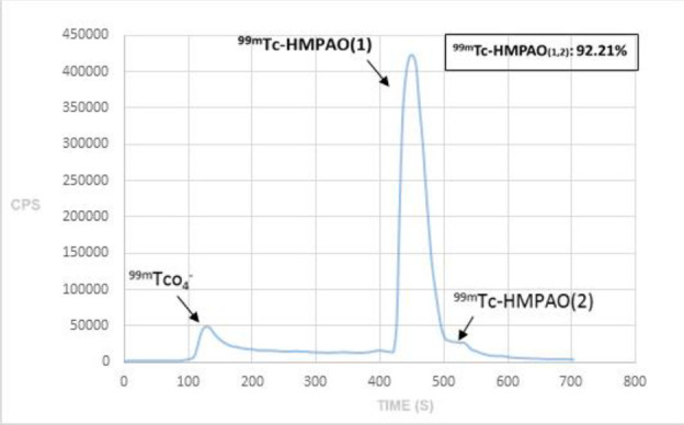
Method: The platelets were extracted from 49 ml whole blood and labeled with Tc-99m HMPAO, then re-injected to the healthy volunteer. Anterior and posterior whole body imaging was done by a dual-head gamma camera 3, 18, 33, 46, 81, 124, 190 min and 15 hours after injection. Also a whole-body SPECT was done at 137 min post-injection. The area under the curves of the spleen, liver, left kidney, bladder, right lung, brain, and abdominal aorta ROIs was calculated to estimate the accumulation of labeled platelets within the organs.
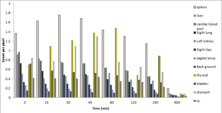
Results: The spleen was the target organ. The kidneys, liver, and heart were also remarkably visualized. The thyroid, stomach, bladder, or gastrointestinal (GI) uptake/activity was not significant. The stomach visualization was enhanced after ingestion at 60 min. The sagittal and lateral sinuses were delineated, and the background of the brain was very low. During the study, the area under the curve of activity was 738, 308, 302, 196, 230, 121, 79, 216, 529, 369, 162, and 54 counts. min/pixel for spleen, liver, heart, right lung, left kidney, right iliac artery, sagittal sinus, thyroid, bladder, stomach, GI, and background, respectively.
Conclusion: The quality of the scan with low dose Tc-99m HMPAO LPs is optimal. We documented the bio-distribution of LPs. The optimal imaging time was 80-120 min post-injection when the free Tc-99m and GI transit were negligible. The sagittal and lateral sinuses were visualized enabling detection of possible clots in the vessels.


 分享
分享
 求助内容:
求助内容: 应助结果提醒方式:
 扫码关注我们
