Pedro F Barbosa, Diego S Gonçalves, Lívia S Ramos, Thaís P Mello, Lys A Braga-Silva, Marcia R Pinto, Carlos P Taborda, Marta H Branquinha, André L S Santos
{"title":"Saps1-3 Antigens in <i>Candida albicans</i>: Differential Modulation Following Exposure to Soluble Proteins, Mammalian Cells, and Infection in Mice.","authors":"Pedro F Barbosa, Diego S Gonçalves, Lívia S Ramos, Thaís P Mello, Lys A Braga-Silva, Marcia R Pinto, Carlos P Taborda, Marta H Branquinha, André L S Santos","doi":"10.3390/idr16040043","DOIUrl":null,"url":null,"abstract":"<p><p>The secreted aspartic peptidases (Saps) of <i>Candida albicans</i> play crucial roles in various steps of fungal-host interactions. Using a flow cytometry approach, this study investigated the expression of Saps1-3 antigens after (i) incubation with soluble proteins, (ii) interaction with mammalian cells, and (iii) infection in immunosuppressed BALB/c mice. Supplementation strategies involving increasing concentrations of bovine serum albumin (BSA) added to yeast carbon base (YCB) medium as the sole nitrogenous source revealed a positive and significant correlation between BSA concentration and both the growth rate and the percentage of fluorescent cells (%FC) labeled with anti-Saps1-3 antibodies. Supplementing the YCB medium with various soluble proteins significantly modulated the expression of Saps1-3 antigens in <i>C. albicans</i>. Specifically, immunoglobulin G, gelatin, and total bovine/human sera significantly reduced the %FC, while laminin, human serum albumin, fibrinogen, hemoglobin, and mucin considerably increased the %FC compared to BSA. Furthermore, co-cultivating <i>C. albicans</i> yeasts with either live epithelial or macrophage cells induced the expression of Saps1-3 antigens in 78% (mean fluorescence intensity [MFI] = 152.1) and 82.7% (MFI = 178.2) of the yeast cells, respectively, compared to BSA, which resulted in 29.3% fluorescent cells (MFI = 50.9). Lastly, the yeasts recovered from the kidneys of infected immunosuppressed mice demonstrated a 4.8-fold increase in the production of Saps1-3 antigens (MFI = 246.6) compared to BSA, with 95.5% of yeasts labeled with anti-Saps1-3 antibodies. Altogether, these results demonstrated the positive modulation of Saps' expression in <i>C. albicans</i> by various key host proteinaceous components, as well as by in vitro and in vivo host challenges.</p>","PeriodicalId":13579,"journal":{"name":"Infectious Disease Reports","volume":"16 4","pages":"572-586"},"PeriodicalIF":2.4000,"publicationDate":"2024-06-28","publicationTypes":"Journal Article","fieldsOfStudy":null,"isOpenAccess":false,"openAccessPdf":"https://www.ncbi.nlm.nih.gov/pmc/articles/PMC11270244/pdf/","citationCount":"0","resultStr":null,"platform":"Semanticscholar","paperid":null,"PeriodicalName":"Infectious Disease Reports","FirstCategoryId":"1085","ListUrlMain":"https://doi.org/10.3390/idr16040043","RegionNum":0,"RegionCategory":null,"ArticlePicture":[],"TitleCN":null,"AbstractTextCN":null,"PMCID":null,"EPubDate":"","PubModel":"","JCR":"Q2","JCRName":"INFECTIOUS DISEASES","Score":null,"Total":0}
引用次数: 0
Abstract
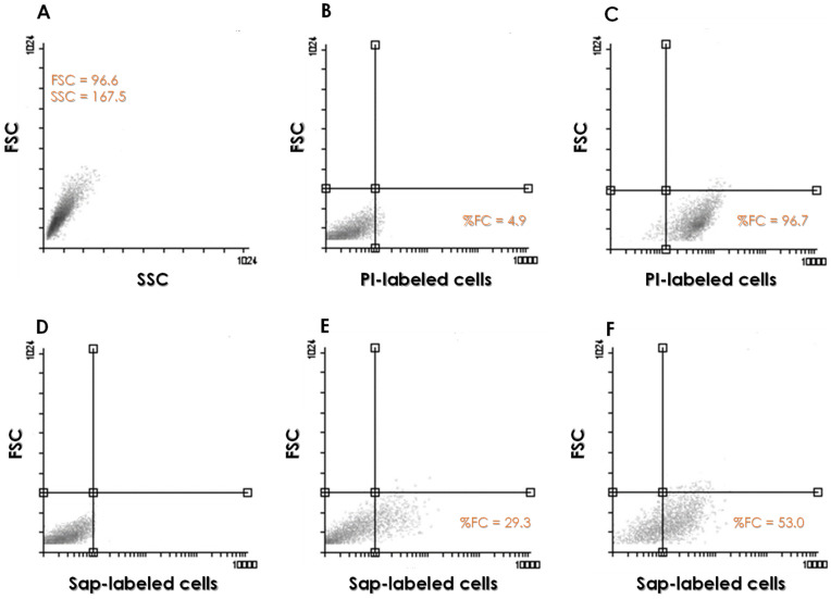
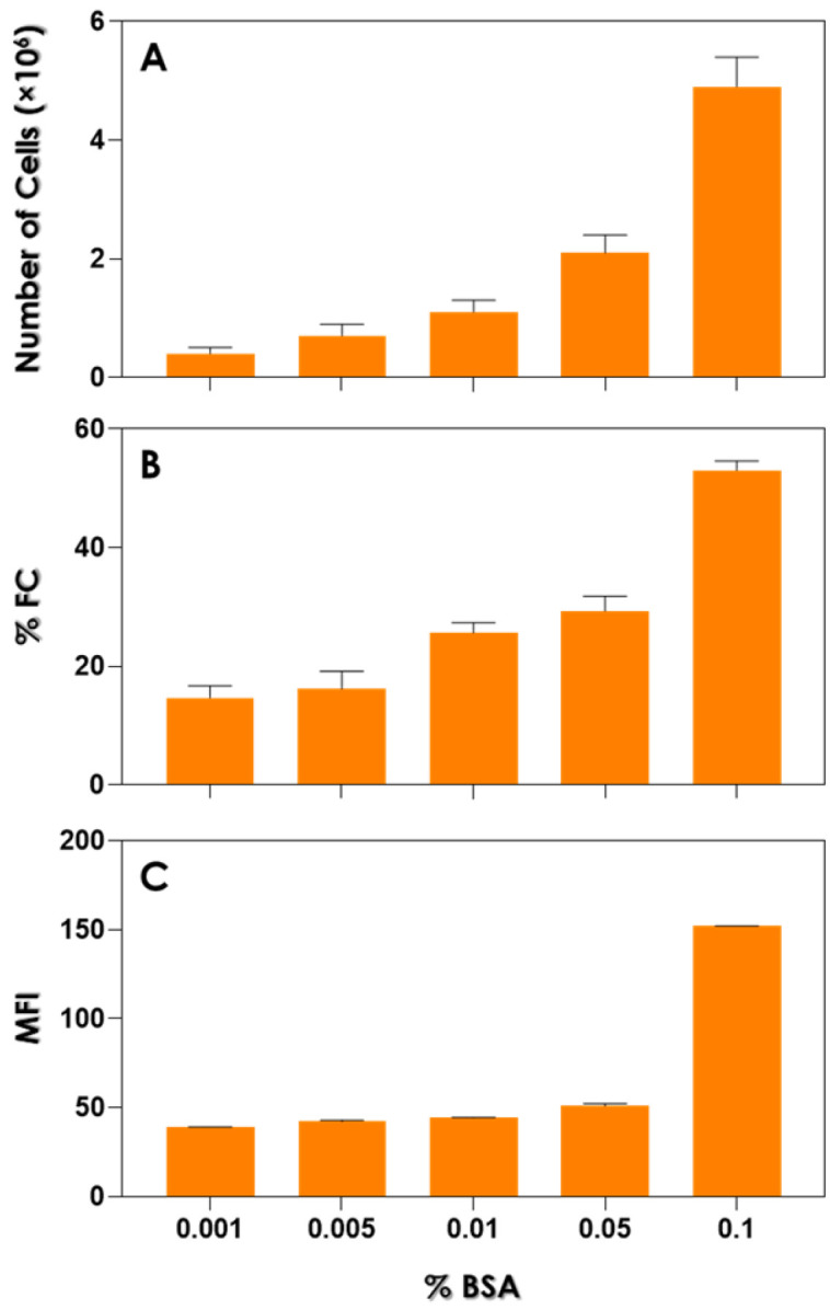
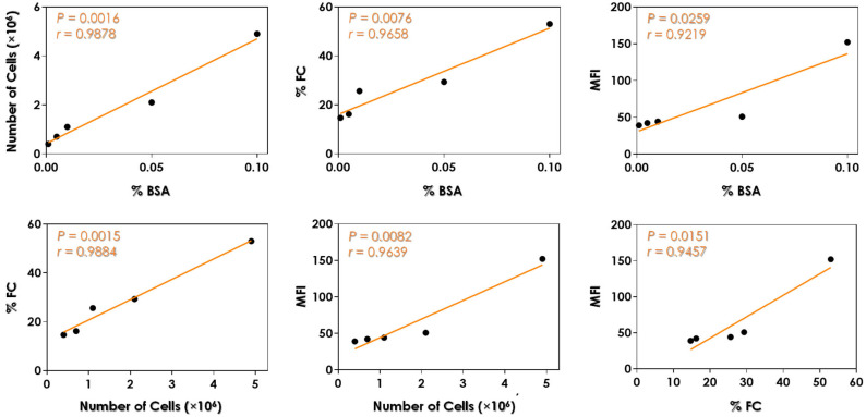
The secreted aspartic peptidases (Saps) of Candida albicans play crucial roles in various steps of fungal-host interactions. Using a flow cytometry approach, this study investigated the expression of Saps1-3 antigens after (i) incubation with soluble proteins, (ii) interaction with mammalian cells, and (iii) infection in immunosuppressed BALB/c mice. Supplementation strategies involving increasing concentrations of bovine serum albumin (BSA) added to yeast carbon base (YCB) medium as the sole nitrogenous source revealed a positive and significant correlation between BSA concentration and both the growth rate and the percentage of fluorescent cells (%FC) labeled with anti-Saps1-3 antibodies. Supplementing the YCB medium with various soluble proteins significantly modulated the expression of Saps1-3 antigens in C. albicans. Specifically, immunoglobulin G, gelatin, and total bovine/human sera significantly reduced the %FC, while laminin, human serum albumin, fibrinogen, hemoglobin, and mucin considerably increased the %FC compared to BSA. Furthermore, co-cultivating C. albicans yeasts with either live epithelial or macrophage cells induced the expression of Saps1-3 antigens in 78% (mean fluorescence intensity [MFI] = 152.1) and 82.7% (MFI = 178.2) of the yeast cells, respectively, compared to BSA, which resulted in 29.3% fluorescent cells (MFI = 50.9). Lastly, the yeasts recovered from the kidneys of infected immunosuppressed mice demonstrated a 4.8-fold increase in the production of Saps1-3 antigens (MFI = 246.6) compared to BSA, with 95.5% of yeasts labeled with anti-Saps1-3 antibodies. Altogether, these results demonstrated the positive modulation of Saps' expression in C. albicans by various key host proteinaceous components, as well as by in vitro and in vivo host challenges.


 分享
分享
 求助内容:
求助内容: 应助结果提醒方式:
 扫码关注我们
