Arada Chaiyamoon, Piyakarn Boontem, Rarinthorn Samrid, Juan J Cardona, Bupachad Khanthiyong, Laphatrada Yurasakpong, Joe Iwanaga, R Shane Tubbs
{"title":"An anatomical study of the nasal foramina.","authors":"Arada Chaiyamoon, Piyakarn Boontem, Rarinthorn Samrid, Juan J Cardona, Bupachad Khanthiyong, Laphatrada Yurasakpong, Joe Iwanaga, R Shane Tubbs","doi":"10.1007/s00276-024-03414-w","DOIUrl":null,"url":null,"abstract":"<p><strong>Purpose: </strong>The nasal foramen is located in the nasal bone and for vessels passage to supply the nasal area. This project aimed to establish reliable references for the nasal foramina for future clinical applications.</p><p><strong>Methods: </strong>The 72 dried skulls, 46 from the Division of Anatomy, University of Phayao, Thailand, and 26 from the Tulane University School of Medicine, USA, were collected and examined. The location, number, and sizes of nasal foramina were noted. The distances from each nasal foramen to the internasal suture, frontonasal suture, nasomaxillary suture, nasion, and rhinion were also recorded and used in the statistical analytical programs.</p><p><strong>Results: </strong>The most common type of nasal foramen in all skulls was type II (one external opening) at 65.97%, followed by type I (no foramen opening) at 20.83%, type III (two external openings) at 11.11% and type IV at 2.08% (three external openings). Nasal foramen subtypes in many of the Thai and American skulls were type IIb and type IIa. The diameter of a connecting nasal foramen was significantly larger than that of a non-connecting. Results from embalmed confirmed the passage of the external nasal artery through the nasal cavity.</p><p><strong>Conclusion: </strong>The study shows no significant difference in nasal foramen morphometry between Thai and American. It illustrates recent data on type and subtype classifications and the location of a vascular passage through the nasal foramen. This is the first study of NF variations and their respective classifications.</p>","PeriodicalId":49461,"journal":{"name":"Surgical and Radiologic Anatomy","volume":" ","pages":"1495-1500"},"PeriodicalIF":1.2000,"publicationDate":"2024-09-01","publicationTypes":"Journal Article","fieldsOfStudy":null,"isOpenAccess":false,"openAccessPdf":"","citationCount":"0","resultStr":null,"platform":"Semanticscholar","paperid":null,"PeriodicalName":"Surgical and Radiologic Anatomy","FirstCategoryId":"3","ListUrlMain":"https://doi.org/10.1007/s00276-024-03414-w","RegionNum":4,"RegionCategory":"医学","ArticlePicture":[],"TitleCN":null,"AbstractTextCN":null,"PMCID":null,"EPubDate":"2024/7/29 0:00:00","PubModel":"Epub","JCR":"Q2","JCRName":"Medicine","Score":null,"Total":0}
引用次数: 0
Abstract
Purpose: The nasal foramen is located in the nasal bone and for vessels passage to supply the nasal area. This project aimed to establish reliable references for the nasal foramina for future clinical applications.
Methods: The 72 dried skulls, 46 from the Division of Anatomy, University of Phayao, Thailand, and 26 from the Tulane University School of Medicine, USA, were collected and examined. The location, number, and sizes of nasal foramina were noted. The distances from each nasal foramen to the internasal suture, frontonasal suture, nasomaxillary suture, nasion, and rhinion were also recorded and used in the statistical analytical programs.
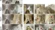
Results: The most common type of nasal foramen in all skulls was type II (one external opening) at 65.97%, followed by type I (no foramen opening) at 20.83%, type III (two external openings) at 11.11% and type IV at 2.08% (three external openings). Nasal foramen subtypes in many of the Thai and American skulls were type IIb and type IIa. The diameter of a connecting nasal foramen was significantly larger than that of a non-connecting. Results from embalmed confirmed the passage of the external nasal artery through the nasal cavity.
Conclusion: The study shows no significant difference in nasal foramen morphometry between Thai and American. It illustrates recent data on type and subtype classifications and the location of a vascular passage through the nasal foramen. This is the first study of NF variations and their respective classifications.
期刊介绍:
Anatomy is a morphological science which cannot fail to interest the clinician. The practical application of anatomical research to clinical problems necessitates special adaptation and selectivity in choosing from numerous international works. Although there is a tendency to believe that meaningful advances in anatomy are unlikely, constant revision is necessary. Surgical and Radiologic Anatomy, the first international journal of Clinical anatomy has been created in this spirit.
Its goal is to serve clinicians, regardless of speciality-physicians, surgeons, radiologists or other specialists-as an indispensable aid with which they can improve their knowledge of anatomy. Each issue includes: Original papers, review articles, articles on the anatomical bases of medical, surgical and radiological techniques, articles of normal radiologic anatomy, brief reviews of anatomical publications of clinical interest.
Particular attention is given to high quality illustrations, which are indispensable for a better understanding of anatomical problems.
Surgical and Radiologic Anatomy is a journal written by anatomists for clinicians with a special interest in anatomy.


 分享
分享
 求助内容:
求助内容: 应助结果提醒方式:
 扫码关注我们
