{"title":"Vagus nerve size determined via ultrasonography is associated with white matter lesions in patients with vascular risk factors.","authors":"Tomohisa Nezu, Futoshi Eto, Akemi Hironaka, Shiro Aoki, Shuichiro Neshige, Saki Tasaka, Hikari Kirimoto, Hirofumi Maruyama","doi":"10.1007/s40477-024-00936-2","DOIUrl":null,"url":null,"abstract":"<p><strong>Purpose: </strong>The cross-sectional area (CSA) of the cervical vagus nerve (VN), as assessed through ultrasonography, might be linked to autonomic nervous system dysfunction. Hypertension is the primary factor associated with cerebral white matter lesions (WMLs), but there is also evidence of a connection with autonomic nervous system dysfunction. However, the associations between WMLs and VN size are unclear. Our objective was to investigate the associations between WMLs and VN size in patients with vascular risk factors.</p><p><strong>Methods: </strong>The CSA of the VN was evaluated using carotid ultrasonography in patients with a history of stroke (acute or chronic) and comorbidities (n = 196, 70.2 ± 12.7 years). Common carotid artery (CCA) intima-media thickness and interadventitial diameter (IAD) were also measured. The severity of the WMLs was assessed by the Fazekas classification and Scheltens' scale.</p><p><strong>Results: </strong>The CSA of the right VN (2.08 ± 0.65 mm<sup>2</sup>) was significantly greater than that of the CSA of the left VN (1.56 ± 0.44 mm<sup>2</sup>) (P < 0.001). Multiple linear regression analyses revealed that older age, hypertension, increased right CCA IAD, and decreased CSA of the right VN (standardized partial regression coefficient [β] - 0.226; P < 0.001) were independently associated with the severity of WMLs (Scheltens' scale). A decreased CSA of the left VN was also associated with the severity of WMLs (β = - 0.239; P < 0.001).</p><p><strong>Conclusion: </strong>VN size determined via ultrasonography was associated with the severity of WMLs. While these findings do not establish a causal relationship, they suggest that autonomic nervous system dysfunction is involved in the progression of WMLs.</p>","PeriodicalId":51528,"journal":{"name":"Journal of Ultrasound","volume":" ","pages":"723-732"},"PeriodicalIF":1.4000,"publicationDate":"2024-09-01","publicationTypes":"Journal Article","fieldsOfStudy":null,"isOpenAccess":false,"openAccessPdf":"https://www.ncbi.nlm.nih.gov/pmc/articles/PMC11333691/pdf/","citationCount":"0","resultStr":null,"platform":"Semanticscholar","paperid":null,"PeriodicalName":"Journal of Ultrasound","FirstCategoryId":"1085","ListUrlMain":"https://doi.org/10.1007/s40477-024-00936-2","RegionNum":0,"RegionCategory":null,"ArticlePicture":[],"TitleCN":null,"AbstractTextCN":null,"PMCID":null,"EPubDate":"2024/7/29 0:00:00","PubModel":"Epub","JCR":"Q3","JCRName":"RADIOLOGY, NUCLEAR MEDICINE & MEDICAL IMAGING","Score":null,"Total":0}
引用次数: 0
Abstract
Purpose: The cross-sectional area (CSA) of the cervical vagus nerve (VN), as assessed through ultrasonography, might be linked to autonomic nervous system dysfunction. Hypertension is the primary factor associated with cerebral white matter lesions (WMLs), but there is also evidence of a connection with autonomic nervous system dysfunction. However, the associations between WMLs and VN size are unclear. Our objective was to investigate the associations between WMLs and VN size in patients with vascular risk factors.
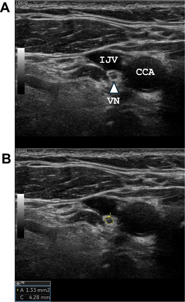
Methods: The CSA of the VN was evaluated using carotid ultrasonography in patients with a history of stroke (acute or chronic) and comorbidities (n = 196, 70.2 ± 12.7 years). Common carotid artery (CCA) intima-media thickness and interadventitial diameter (IAD) were also measured. The severity of the WMLs was assessed by the Fazekas classification and Scheltens' scale.
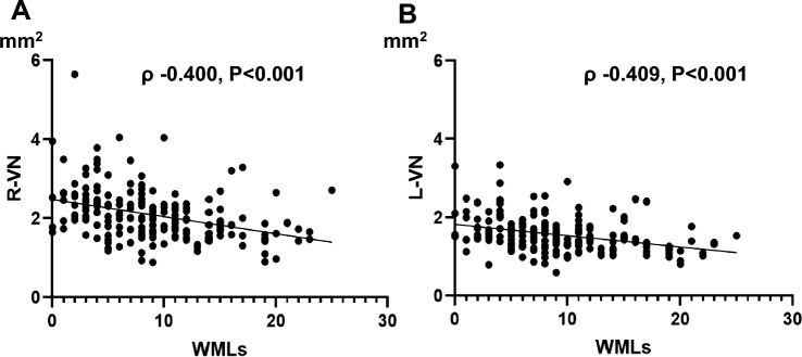
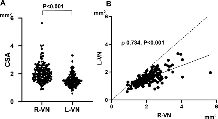
Results: The CSA of the right VN (2.08 ± 0.65 mm2) was significantly greater than that of the CSA of the left VN (1.56 ± 0.44 mm2) (P < 0.001). Multiple linear regression analyses revealed that older age, hypertension, increased right CCA IAD, and decreased CSA of the right VN (standardized partial regression coefficient [β] - 0.226; P < 0.001) were independently associated with the severity of WMLs (Scheltens' scale). A decreased CSA of the left VN was also associated with the severity of WMLs (β = - 0.239; P < 0.001).
Conclusion: VN size determined via ultrasonography was associated with the severity of WMLs. While these findings do not establish a causal relationship, they suggest that autonomic nervous system dysfunction is involved in the progression of WMLs.
期刊介绍:
The Journal of Ultrasound is the official journal of the Italian Society for Ultrasound in Medicine and Biology (SIUMB). The journal publishes original contributions (research and review articles, case reports, technical reports and letters to the editor) on significant advances in clinical diagnostic, interventional and therapeutic applications, clinical techniques, the physics, engineering and technology of ultrasound in medicine and biology, and in cross-sectional diagnostic imaging. The official language of Journal of Ultrasound is English.


 分享
分享
 求助内容:
求助内容: 应助结果提醒方式:
 扫码关注我们
