Basil E Grüter, Kristina Catalano, Javier Anon, Philipp Gruber, Arjun Thanabalasingam, Lukas Andereggen, Gerrit A Schubert, Luca Remonda, Serge Marbacher
{"title":"Intra-aneurysmal contrast agent stasis during intraoperative digital subtraction angiography may predict long-term occlusion after clipping.","authors":"Basil E Grüter, Kristina Catalano, Javier Anon, Philipp Gruber, Arjun Thanabalasingam, Lukas Andereggen, Gerrit A Schubert, Luca Remonda, Serge Marbacher","doi":"10.1007/s00701-024-06162-3","DOIUrl":null,"url":null,"abstract":"<p><strong>Purpose: </strong>The routine use of intraoperative digital subtraction angiography (iDSA) increases detection of intracranial aneurysm (IA) remnants after microsurgical clipping. Spontaneous thrombosis of IA remnants after clipping is considered a rare phenomenon. We analyse iDSA characteristics to find predictors for IA remnant thrombosis.</p><p><strong>Methods: </strong>IA with intraoperative detection of a remnant after clipping were identified and divided into remnants experiencing spontaneous thrombosis, and remnants with long-term patency and/or remnant growth. Angiographic features of iDSA were analysed and compared between the two groups.</p><p><strong>Results: </strong>Of 37 IAs with intraoperative remnant on 3D-iDSA, five sustained a spontaneous remnant thrombosis and remained occluded in long-term follow-up. In all five cases, iDSA revealed delayed inflow and consequent stasis of the contrast agent until the late venous phase. On the other hand, in all cases with persistent long-term IA remnants (n = 32) iDSA demonstrated timely arterial contrast inflow and wash-out without stasis of intra-aneurysmal contrast agent.</p><p><strong>Conclusions: </strong>Contrast stasis in IA remnants during iDSA appears to predict long-term IA occlusion, indicating that clip correction manoeuvres or even attempted endovascular treatment of the remnant IA may be avoided in these patients.</p>","PeriodicalId":7370,"journal":{"name":"Acta Neurochirurgica","volume":"166 1","pages":"309"},"PeriodicalIF":1.9000,"publicationDate":"2024-07-30","publicationTypes":"Journal Article","fieldsOfStudy":null,"isOpenAccess":false,"openAccessPdf":"","citationCount":"0","resultStr":null,"platform":"Semanticscholar","paperid":null,"PeriodicalName":"Acta Neurochirurgica","FirstCategoryId":"3","ListUrlMain":"https://doi.org/10.1007/s00701-024-06162-3","RegionNum":3,"RegionCategory":"医学","ArticlePicture":[],"TitleCN":null,"AbstractTextCN":null,"PMCID":null,"EPubDate":"","PubModel":"","JCR":"Q3","JCRName":"CLINICAL NEUROLOGY","Score":null,"Total":0}
引用次数: 0
Abstract
Purpose: The routine use of intraoperative digital subtraction angiography (iDSA) increases detection of intracranial aneurysm (IA) remnants after microsurgical clipping. Spontaneous thrombosis of IA remnants after clipping is considered a rare phenomenon. We analyse iDSA characteristics to find predictors for IA remnant thrombosis.
Methods: IA with intraoperative detection of a remnant after clipping were identified and divided into remnants experiencing spontaneous thrombosis, and remnants with long-term patency and/or remnant growth. Angiographic features of iDSA were analysed and compared between the two groups.
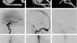
Results: Of 37 IAs with intraoperative remnant on 3D-iDSA, five sustained a spontaneous remnant thrombosis and remained occluded in long-term follow-up. In all five cases, iDSA revealed delayed inflow and consequent stasis of the contrast agent until the late venous phase. On the other hand, in all cases with persistent long-term IA remnants (n = 32) iDSA demonstrated timely arterial contrast inflow and wash-out without stasis of intra-aneurysmal contrast agent.
Conclusions: Contrast stasis in IA remnants during iDSA appears to predict long-term IA occlusion, indicating that clip correction manoeuvres or even attempted endovascular treatment of the remnant IA may be avoided in these patients.
期刊介绍:
The journal "Acta Neurochirurgica" publishes only original papers useful both to research and clinical work. Papers should deal with clinical neurosurgery - diagnosis and diagnostic techniques, operative surgery and results, postoperative treatment - or with research work in neuroscience if the underlying questions or the results are of neurosurgical interest. Reports on congresses are given in brief accounts. As official organ of the European Association of Neurosurgical Societies the journal publishes all announcements of the E.A.N.S. and reports on the activities of its member societies. Only contributions written in English will be accepted.


 分享
分享
 求助内容:
求助内容: 应助结果提醒方式:
 扫码关注我们
