Busra Ozturk, Yavuz Selim Taspinar, Murat Koklu, Melek Tassoker
{"title":"Automatic segmentation of the maxillary sinus on cone beam computed tomographic images with U-Net deep learning model.","authors":"Busra Ozturk, Yavuz Selim Taspinar, Murat Koklu, Melek Tassoker","doi":"10.1007/s00405-024-08870-z","DOIUrl":null,"url":null,"abstract":"<p><strong>Background: </strong>Medical imaging segmentation is the use of image processing techniques to expand specific structures or areas in medical images. This technique is used to separate and display different textures or shapes in an image. The aim of this study is to develop a deep learning-based method to perform maxillary sinus segmentation using cone beam computed tomography (CBCT) images. The proposed segmentation method aims to provide better image guidance to surgeons and specialists by determining the boundaries of the maxillary sinus cavities. In this way, more accurate diagnoses can be made and surgical interventions can be performed more successfully.</p><p><strong>Methods: </strong>In the study, axial CBCT images of 100 patients (200 maxillary sinuses) were used. These images were marked to identify the maxillary sinus walls. The marked regions are masked for use in the maxillary sinus segmentation model. U-Net, one of the deep learning methods, was used for segmentation. The training process was carried out for 10 epochs and 100 iterations per epoch. The epoch and iteration numbers in which the model showed maximum success were determined using the early stopping method.</p><p><strong>Results: </strong>After the segmentation operations performed with the U-Net model trained using CBCT images, both visual and numerical results were obtained. In order to measure the performance of the U-Net model, IoU (Intersection over Union) and F1 Score metrics were used. As a result of the tests of the model, the IoU value was found to be 0.9275 and the F1 Score value was 0.9784.</p><p><strong>Conclusion: </strong>The U-Net model has shown high success in maxillary sinus segmentation. In this way, fast and highly accurate evaluations are possible, saving time by reducing the workload of clinicians and eliminating subjective errors.</p>","PeriodicalId":11952,"journal":{"name":"European Archives of Oto-Rhino-Laryngology","volume":" ","pages":"6111-6121"},"PeriodicalIF":2.2000,"publicationDate":"2024-11-01","publicationTypes":"Journal Article","fieldsOfStudy":null,"isOpenAccess":false,"openAccessPdf":"https://www.ncbi.nlm.nih.gov/pmc/articles/PMC11512868/pdf/","citationCount":"0","resultStr":null,"platform":"Semanticscholar","paperid":null,"PeriodicalName":"European Archives of Oto-Rhino-Laryngology","FirstCategoryId":"3","ListUrlMain":"https://doi.org/10.1007/s00405-024-08870-z","RegionNum":3,"RegionCategory":"医学","ArticlePicture":[],"TitleCN":null,"AbstractTextCN":null,"PMCID":null,"EPubDate":"2024/7/31 0:00:00","PubModel":"Epub","JCR":"Q2","JCRName":"OTORHINOLARYNGOLOGY","Score":null,"Total":0}
引用次数: 0
Abstract
Background: Medical imaging segmentation is the use of image processing techniques to expand specific structures or areas in medical images. This technique is used to separate and display different textures or shapes in an image. The aim of this study is to develop a deep learning-based method to perform maxillary sinus segmentation using cone beam computed tomography (CBCT) images. The proposed segmentation method aims to provide better image guidance to surgeons and specialists by determining the boundaries of the maxillary sinus cavities. In this way, more accurate diagnoses can be made and surgical interventions can be performed more successfully.
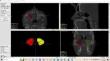
Methods: In the study, axial CBCT images of 100 patients (200 maxillary sinuses) were used. These images were marked to identify the maxillary sinus walls. The marked regions are masked for use in the maxillary sinus segmentation model. U-Net, one of the deep learning methods, was used for segmentation. The training process was carried out for 10 epochs and 100 iterations per epoch. The epoch and iteration numbers in which the model showed maximum success were determined using the early stopping method.
Results: After the segmentation operations performed with the U-Net model trained using CBCT images, both visual and numerical results were obtained. In order to measure the performance of the U-Net model, IoU (Intersection over Union) and F1 Score metrics were used. As a result of the tests of the model, the IoU value was found to be 0.9275 and the F1 Score value was 0.9784.
Conclusion: The U-Net model has shown high success in maxillary sinus segmentation. In this way, fast and highly accurate evaluations are possible, saving time by reducing the workload of clinicians and eliminating subjective errors.
期刊介绍:
Official Journal of
European Union of Medical Specialists – ORL Section and Board
Official Journal of Confederation of European Oto-Rhino-Laryngology Head and Neck Surgery
"European Archives of Oto-Rhino-Laryngology" publishes original clinical reports and clinically relevant experimental studies, as well as short communications presenting new results of special interest. With peer review by a respected international editorial board and prompt English-language publication, the journal provides rapid dissemination of information by authors from around the world. This particular feature makes it the journal of choice for readers who want to be informed about the continuing state of the art concerning basic sciences and the diagnosis and management of diseases of the head and neck on an international level.
European Archives of Oto-Rhino-Laryngology was founded in 1864 as "Archiv für Ohrenheilkunde" by A. von Tröltsch, A. Politzer and H. Schwartze.


 分享
分享
 求助内容:
求助内容: 应助结果提醒方式:
 扫码关注我们
