{"title":"CTSG may inhibit disease progression in HIV-related lung cancer patients by affecting immunosuppression.","authors":"Xuan Yan, Shuoyan Wei, Yuexiang Yang, Zhangyan Zhao, Qingguo Wu, Haicheng Tang","doi":"10.1186/s13027-024-00599-y","DOIUrl":null,"url":null,"abstract":"<p><strong>Objectives: </strong>Lung cancer is an independent risk factor for pulmonary complications following HIV infection. This study aimed to examine the expression and clinical significance of Cathepsin G (CTSG) protein in both non-HIV and HIV-related lung cancers.</p><p><strong>Methods: </strong>The data related to lung adenocarcinoma (LUAD) and lung squamous carcinoma (LUSC) in the TCGA dataset and the data related to healthy individuals in the GTEx dataset, the GEPIA2 database was used to excavate the distinction in the expression of CTSG protein in non-small cell lung cancer (NSCLC) tissues versus normal non-cancerous tissues. The Ualcan database was used to compare the differences in CTSG expression at different stages of LUAD and LUSC. Immunohistochemistry (IHC) was used to detect the expression of CTSG proteins in the pathological tissues of patients with HIV-related lung cancer and patients with lung cancer without co-infection, the Kaplan-Meier method was used for survival analysis.</p><p><strong>Results: </strong>We observed that CTSG expression in NSCLC is lower compared to adjacent non-tumor tissues and correlates with NSCLC clinical stage. CTSG protein expression in HIV-related lung cancer tissues was lower than in adjacent tissues and lower than in lung cancer tissues without HIV infection, with a statistically significant difference (P < 0.05). It correlated with CD4 + T cell count and CD4+/CD8 + T cell ratio, as well as with the pathological type, distant metastasis, and clinical stage of HIV-related lung cancer, all with statistical significance (P < 0.05).</p><p><strong>Conclusions: </strong>CTSG could potentially mitigate disease advancement in HIV-related lung cancer patients by inhibiting immune depletion, serving as a prospective immunotherapeutic target for both non-HIV and HIV-associated lung cancers.</p>","PeriodicalId":13568,"journal":{"name":"Infectious Agents and Cancer","volume":"19 1","pages":"34"},"PeriodicalIF":2.8000,"publicationDate":"2024-07-30","publicationTypes":"Journal Article","fieldsOfStudy":null,"isOpenAccess":false,"openAccessPdf":"https://www.ncbi.nlm.nih.gov/pmc/articles/PMC11290089/pdf/","citationCount":"0","resultStr":null,"platform":"Semanticscholar","paperid":null,"PeriodicalName":"Infectious Agents and Cancer","FirstCategoryId":"3","ListUrlMain":"https://doi.org/10.1186/s13027-024-00599-y","RegionNum":2,"RegionCategory":"医学","ArticlePicture":[],"TitleCN":null,"AbstractTextCN":null,"PMCID":null,"EPubDate":"","PubModel":"","JCR":"Q3","JCRName":"IMMUNOLOGY","Score":null,"Total":0}
引用次数: 0
Abstract
Objectives: Lung cancer is an independent risk factor for pulmonary complications following HIV infection. This study aimed to examine the expression and clinical significance of Cathepsin G (CTSG) protein in both non-HIV and HIV-related lung cancers.
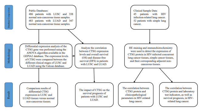
Methods: The data related to lung adenocarcinoma (LUAD) and lung squamous carcinoma (LUSC) in the TCGA dataset and the data related to healthy individuals in the GTEx dataset, the GEPIA2 database was used to excavate the distinction in the expression of CTSG protein in non-small cell lung cancer (NSCLC) tissues versus normal non-cancerous tissues. The Ualcan database was used to compare the differences in CTSG expression at different stages of LUAD and LUSC. Immunohistochemistry (IHC) was used to detect the expression of CTSG proteins in the pathological tissues of patients with HIV-related lung cancer and patients with lung cancer without co-infection, the Kaplan-Meier method was used for survival analysis.
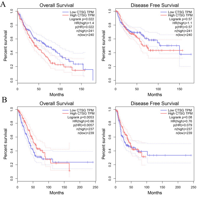
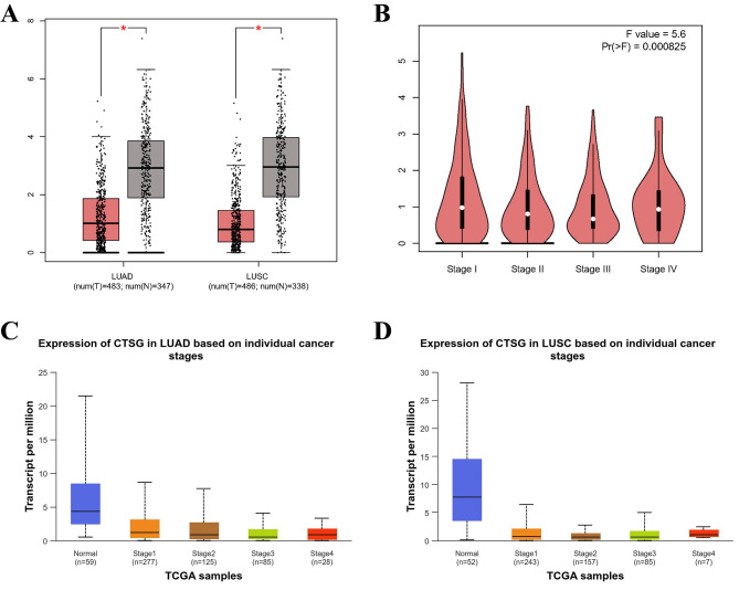
Results: We observed that CTSG expression in NSCLC is lower compared to adjacent non-tumor tissues and correlates with NSCLC clinical stage. CTSG protein expression in HIV-related lung cancer tissues was lower than in adjacent tissues and lower than in lung cancer tissues without HIV infection, with a statistically significant difference (P < 0.05). It correlated with CD4 + T cell count and CD4+/CD8 + T cell ratio, as well as with the pathological type, distant metastasis, and clinical stage of HIV-related lung cancer, all with statistical significance (P < 0.05).
Conclusions: CTSG could potentially mitigate disease advancement in HIV-related lung cancer patients by inhibiting immune depletion, serving as a prospective immunotherapeutic target for both non-HIV and HIV-associated lung cancers.
期刊介绍:
Infectious Agents and Cancer is an open access, peer-reviewed online journal that encompasses all aspects of basic, clinical, epidemiological and translational research providing an insight into the association between chronic infections and cancer.
The journal welcomes submissions in the pathogen-related cancer areas and other related topics, in particular:
• HPV and anogenital cancers, as well as head and neck cancers;
• EBV and Burkitt lymphoma;
• HCV/HBV and hepatocellular carcinoma as well as lymphoproliferative diseases;
• HHV8 and Kaposi sarcoma;
• HTLV and leukemia;
• Cancers in Low- and Middle-income countries.
The link between infection and cancer has become well established over the past 50 years, and infection-associated cancer contribute up to 16% of cancers in developed countries and 33% in less developed countries.
Preventive vaccines have been developed for only two cancer-causing viruses, highlighting both the opportunity to prevent infection-associated cancers by vaccination and the gaps that remain before vaccines can be developed for other cancer-causing agents. These gaps are due to incomplete understanding of the basic biology, natural history, epidemiology of many of the pathogens that cause cancer, the mechanisms they exploit to cause cancer, and how to interrupt progression to cancer in human populations. Early diagnosis or identification of lesions at high risk of progression represent the current most critical research area of the field supported by recent advances in genomics and proteomics technologies.


 分享
分享
 求助内容:
求助内容: 应助结果提醒方式:
 扫码关注我们
