Dan Alexandru Cozac, Eleonora Lassandro, Raffaella Motta, Valeria Pergola
{"title":"Caseous Calcification of the Mitral Annulus Associated with Severe Mitral Regurgitation: A Multimodality Diagnostic Approach.","authors":"Dan Alexandru Cozac, Eleonora Lassandro, Raffaella Motta, Valeria Pergola","doi":"10.4103/jcecho.jcecho_20_24","DOIUrl":null,"url":null,"abstract":"<p><p>Caseous calcification of the mitral annulus (CCMA) is a rare variant of mitral annular calcification, and a multimodality approach is advised to ensure an accurate diagnosis. We report a case of a patient with CCMA, associated with severe mitral regurgitation. An 82-year-old woman was admitted due to worsening heart failure. Transthoracic echocardiography revealed a fixed, hyperechogenic mass, accompanied by restriction of the posterior mitral leaflet, and subsequent severe mitral regurgitation. Transesophageal echocardiography demonstrated a restricted motion of the posterior mitral leaflet, because of a large, echogenic mass (15 mm × 11 mm), attached to the mitral annulus, vacuolated with a central echolucent aspect, lacking acoustic shadowing. Contrast-enhanced cardiac computed tomography identified a distinct oval mass (18 mm × 11 mm × 19 mm) presenting a central hypodense content and peripheral calcification, strongly suggestive of CCMA. Considering the patient's profile, surgical valvular replacement was considered unsuitable. Therefore, a transcatheter edge-to-edge repair was performed, resulting in mild residual regurgitation.</p>","PeriodicalId":15191,"journal":{"name":"Journal of Cardiovascular Echography","volume":"34 2","pages":"82-84"},"PeriodicalIF":1.0000,"publicationDate":"2024-04-01","publicationTypes":"Journal Article","fieldsOfStudy":null,"isOpenAccess":false,"openAccessPdf":"https://www.ncbi.nlm.nih.gov/pmc/articles/PMC11288304/pdf/","citationCount":"0","resultStr":null,"platform":"Semanticscholar","paperid":null,"PeriodicalName":"Journal of Cardiovascular Echography","FirstCategoryId":"1085","ListUrlMain":"https://doi.org/10.4103/jcecho.jcecho_20_24","RegionNum":0,"RegionCategory":null,"ArticlePicture":[],"TitleCN":null,"AbstractTextCN":null,"PMCID":null,"EPubDate":"2024/6/28 0:00:00","PubModel":"Epub","JCR":"Q4","JCRName":"CARDIAC & CARDIOVASCULAR SYSTEMS","Score":null,"Total":0}
引用次数: 0
Abstract
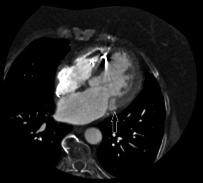
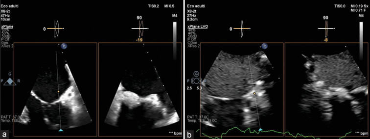
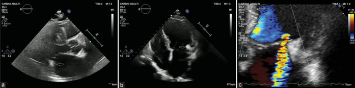
Caseous calcification of the mitral annulus (CCMA) is a rare variant of mitral annular calcification, and a multimodality approach is advised to ensure an accurate diagnosis. We report a case of a patient with CCMA, associated with severe mitral regurgitation. An 82-year-old woman was admitted due to worsening heart failure. Transthoracic echocardiography revealed a fixed, hyperechogenic mass, accompanied by restriction of the posterior mitral leaflet, and subsequent severe mitral regurgitation. Transesophageal echocardiography demonstrated a restricted motion of the posterior mitral leaflet, because of a large, echogenic mass (15 mm × 11 mm), attached to the mitral annulus, vacuolated with a central echolucent aspect, lacking acoustic shadowing. Contrast-enhanced cardiac computed tomography identified a distinct oval mass (18 mm × 11 mm × 19 mm) presenting a central hypodense content and peripheral calcification, strongly suggestive of CCMA. Considering the patient's profile, surgical valvular replacement was considered unsuitable. Therefore, a transcatheter edge-to-edge repair was performed, resulting in mild residual regurgitation.


 分享
分享
 求助内容:
求助内容: 应助结果提醒方式:
 扫码关注我们
