Francesca Silvestrini-Biavati, Giorgio Oliva, Luis Huanca Ghislanzoni, Elisa Ottonelli, Domenico Dalessandri, Valentina Lanteri, Alessandro Ugolini
{"title":"Evaluation of palate morphology in patients treated with leaf expander and hyrax expander: A geometric morphometric analysis","authors":"Francesca Silvestrini-Biavati, Giorgio Oliva, Luis Huanca Ghislanzoni, Elisa Ottonelli, Domenico Dalessandri, Valentina Lanteri, Alessandro Ugolini","doi":"10.1111/ocr.12839","DOIUrl":null,"url":null,"abstract":"<div>\n \n \n <section>\n \n <h3> Objective</h3>\n \n <p>The aim of this study was to evaluate changes in shape of the palatal vault after maxillary expansion with hyrax expander (HE) and leaf expander (LE), using 3D Geometric Morphometric Analysis.</p>\n </section>\n \n <section>\n \n <h3> Setting and Sample Population</h3>\n \n <p>Overall, 250 patients (110 M, 140 F) with maxillary transverse deficiency were selected for this study. In this study, 127 subjects were treated with HE, 123 with LE.</p>\n </section>\n \n <section>\n \n <h3> Materials and Methods</h3>\n \n <p>Digital dental models were obtained pre-treatment (T0) and after 12 months from the cementation of the device (T1) and processed by means of a digital scanner. Linear and morphometric analyses were conducted to determine the effects of each appliance on dental measurements and palatal shape, and a multiple linear regression was performed to analyse the influence of anchorage and appliance type on final shape.</p>\n </section>\n \n <section>\n \n <h3> Results</h3>\n \n <p>Morphometric analysis showed that there was a lowering of the palatal vault in the HE group, while in the LE group it remained unchanged: the difference in palatal shape at time T0 and T1 was statistically significant in both treatments (HE vs. LE). In the HE group, the change in shape also included the upper part of the palatal vault in the vertical dimension, while in the LE group the change in shape interested mainly palatal shelves and the lower portion of the palate.</p>\n </section>\n \n <section>\n \n <h3> Conclusions</h3>\n \n <p>Both LE and HE produce clinically significant changes in the morphology of the palatal vault.</p>\n </section>\n </div>","PeriodicalId":19652,"journal":{"name":"Orthodontics & Craniofacial Research","volume":"27 6","pages":"959-966"},"PeriodicalIF":1.7000,"publicationDate":"2024-08-02","publicationTypes":"Journal Article","fieldsOfStudy":null,"isOpenAccess":false,"openAccessPdf":"https://onlinelibrary.wiley.com/doi/epdf/10.1111/ocr.12839","citationCount":"0","resultStr":null,"platform":"Semanticscholar","paperid":null,"PeriodicalName":"Orthodontics & Craniofacial Research","FirstCategoryId":"3","ListUrlMain":"https://onlinelibrary.wiley.com/doi/10.1111/ocr.12839","RegionNum":3,"RegionCategory":"医学","ArticlePicture":[],"TitleCN":null,"AbstractTextCN":null,"PMCID":null,"EPubDate":"","PubModel":"","JCR":"Q2","JCRName":"DENTISTRY, ORAL SURGERY & MEDICINE","Score":null,"Total":0}
引用次数: 0
Abstract
Objective
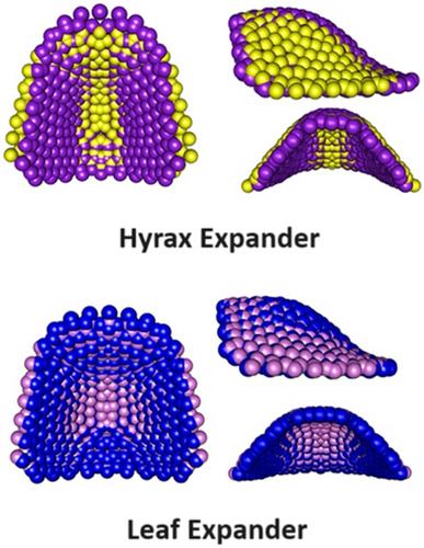
The aim of this study was to evaluate changes in shape of the palatal vault after maxillary expansion with hyrax expander (HE) and leaf expander (LE), using 3D Geometric Morphometric Analysis.
Setting and Sample Population
Overall, 250 patients (110 M, 140 F) with maxillary transverse deficiency were selected for this study. In this study, 127 subjects were treated with HE, 123 with LE.
Materials and Methods
Digital dental models were obtained pre-treatment (T0) and after 12 months from the cementation of the device (T1) and processed by means of a digital scanner. Linear and morphometric analyses were conducted to determine the effects of each appliance on dental measurements and palatal shape, and a multiple linear regression was performed to analyse the influence of anchorage and appliance type on final shape.
Results
Morphometric analysis showed that there was a lowering of the palatal vault in the HE group, while in the LE group it remained unchanged: the difference in palatal shape at time T0 and T1 was statistically significant in both treatments (HE vs. LE). In the HE group, the change in shape also included the upper part of the palatal vault in the vertical dimension, while in the LE group the change in shape interested mainly palatal shelves and the lower portion of the palate.
Conclusions
Both LE and HE produce clinically significant changes in the morphology of the palatal vault.
期刊介绍:
Orthodontics & Craniofacial Research - Genes, Growth and Development is published to serve its readers as an international forum for the presentation and critical discussion of issues pertinent to the advancement of the specialty of orthodontics and the evidence-based knowledge of craniofacial growth and development. This forum is based on scientifically supported information, but also includes minority and conflicting opinions.
The objective of the journal is to facilitate effective communication between the research community and practicing clinicians. Original papers of high scientific quality that report the findings of clinical trials, clinical epidemiology, and novel therapeutic or diagnostic approaches are appropriate submissions. Similarly, we welcome papers in genetics, developmental biology, syndromology, surgery, speech and hearing, and other biomedical disciplines related to clinical orthodontics and normal and abnormal craniofacial growth and development. In addition to original and basic research, the journal publishes concise reviews, case reports of substantial value, invited essays, letters, and announcements.
The journal is published quarterly. The review of submitted papers will be coordinated by the editor and members of the editorial board. It is policy to review manuscripts within 3 to 4 weeks of receipt and to publish within 3 to 6 months of acceptance.


 分享
分享
 求助内容:
求助内容: 应助结果提醒方式:
 扫码关注我们
