S. Greisert, S. Fleck, E. Rathmann, M. Vollmer, H. W. S. Schroeder
{"title":"The role of MRI biomarkers in evaluation of symptomatic pineal cysts – a retrospective analysis","authors":"S. Greisert, S. Fleck, E. Rathmann, M. Vollmer, H. W. S. Schroeder","doi":"10.1007/s00701-024-06212-w","DOIUrl":null,"url":null,"abstract":"<h3 data-test=\"abstract-sub-heading\">Background</h3><p>Our aim was to determine whether the Apparent Diffusion Coefficient is able to predict the presence of a symptomatic pineal cyst by detecting cerebral edema.</p><h3 data-test=\"abstract-sub-heading\">Methods</h3><p>We retrospectively analyzed MRIs of 45 patients with pineal cysts before and after resection and 51 patients without pineal cysts, comparing ADC values of thalamus, central, periventricular and subcortical white matter. Furthermore we evaluated cyst size and morphology and analyzed its correlation to ADC values in corresponding patients.</p><h3 data-test=\"abstract-sub-heading\">Results</h3><p>Differences between patients with symptomatic pineal cyst and control group were not significant (<i>p</i> = 0.200 – 0.968). ADC ratios did not change significantly after resection of the cyst (<i>p</i> = 0.575 – 0.862). Cyst size showed no significant correlation to ADC ratios (<i>p</i> = 0.071 – 0.918). Raw data analyses revealed more significance, especially periventricularly and in central white matter, which resulted in significant interhemispheric differences in ADC ratios in both subgroups (<i>p</i> < 0.001 and <i>p</i> = 0.031). MRI of 1.5T showed consistently higher values than 3T but mostly insignificant.</p><h3 data-test=\"abstract-sub-heading\">Conclusion</h3><p>Our analysis revealed no evidence that pineal cysts lead to intracerebral edema caused by venous compression. Since variability was higher than the differences seen, ADC sequences do not appear to be an appropriate diagnostic tool for symptomatic pineal cysts.</p>","PeriodicalId":7370,"journal":{"name":"Acta Neurochirurgica","volume":"35 1","pages":""},"PeriodicalIF":1.9000,"publicationDate":"2024-08-03","publicationTypes":"Journal Article","fieldsOfStudy":null,"isOpenAccess":false,"openAccessPdf":"","citationCount":"0","resultStr":null,"platform":"Semanticscholar","paperid":null,"PeriodicalName":"Acta Neurochirurgica","FirstCategoryId":"3","ListUrlMain":"https://doi.org/10.1007/s00701-024-06212-w","RegionNum":3,"RegionCategory":"医学","ArticlePicture":[],"TitleCN":null,"AbstractTextCN":null,"PMCID":null,"EPubDate":"","PubModel":"","JCR":"Q3","JCRName":"CLINICAL NEUROLOGY","Score":null,"Total":0}
引用次数: 0
Abstract
Background
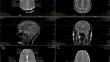
Our aim was to determine whether the Apparent Diffusion Coefficient is able to predict the presence of a symptomatic pineal cyst by detecting cerebral edema.
Methods
We retrospectively analyzed MRIs of 45 patients with pineal cysts before and after resection and 51 patients without pineal cysts, comparing ADC values of thalamus, central, periventricular and subcortical white matter. Furthermore we evaluated cyst size and morphology and analyzed its correlation to ADC values in corresponding patients.
Results
Differences between patients with symptomatic pineal cyst and control group were not significant (p = 0.200 – 0.968). ADC ratios did not change significantly after resection of the cyst (p = 0.575 – 0.862). Cyst size showed no significant correlation to ADC ratios (p = 0.071 – 0.918). Raw data analyses revealed more significance, especially periventricularly and in central white matter, which resulted in significant interhemispheric differences in ADC ratios in both subgroups (p < 0.001 and p = 0.031). MRI of 1.5T showed consistently higher values than 3T but mostly insignificant.
Conclusion
Our analysis revealed no evidence that pineal cysts lead to intracerebral edema caused by venous compression. Since variability was higher than the differences seen, ADC sequences do not appear to be an appropriate diagnostic tool for symptomatic pineal cysts.
期刊介绍:
The journal "Acta Neurochirurgica" publishes only original papers useful both to research and clinical work. Papers should deal with clinical neurosurgery - diagnosis and diagnostic techniques, operative surgery and results, postoperative treatment - or with research work in neuroscience if the underlying questions or the results are of neurosurgical interest. Reports on congresses are given in brief accounts. As official organ of the European Association of Neurosurgical Societies the journal publishes all announcements of the E.A.N.S. and reports on the activities of its member societies. Only contributions written in English will be accepted.


 分享
分享
 求助内容:
求助内容: 应助结果提醒方式:
 扫码关注我们
