{"title":"A novel electroencephalographic evaluation of noxious stimulation during isoflurane anesthesia in dogs.","authors":"Wei-Mao Hung, Hsien-Chi Wang, Julia Chu-Ning Hsu","doi":"10.1538/expanim.24-0036","DOIUrl":null,"url":null,"abstract":"<p><p>In veterinary clinical medicine, evaluating the balance between nociception and antinociception presents a great challenge for anesthesiologists during canine surgeries. Heart rate (HR) and mean arterial pressure (MAP) are suitable indexes for monitoring noxious stimuli during anesthesia. Frontal electroencephalography (EEG) records, including processed parameters, are recommended for evaluating nociceptive balance in anesthetized unconscious human patients, which is unexplored in veterinary medicine. Therefore, the objective is to explore the response of processed EEG parameters to noxious stimulation and elucidate the impact of noxious stimulation on frontal cortical activity in dogs anesthetized with 1.5% isoflurane. Fourteen dogs were included and underwent frontal EEG monitoring, measuring the patient state index (PSI) and spectral edge frequency (SEF) before and after administering noxious stimulation using the towel clamp method on the tail of each 1.5% isoflurane-anesthetized dog. As the noxious stimulation was applied, there was a simultaneous increase in PSI, HR, and MAP, with PSI exhibiting a drastic response. SEF, especially on the left side, also increased with noxious stimulation. In EEG power spectral analysis, the delta band was decreased, and the alpha and beta bands showed an increase following noxious stimulation, with a more profound elevation of beta band on the left side. This study suggests that noxious stimulation brings asymmetric frontal cortical arousal, changing brain activity by suppressing delta wave and augmenting alpha and beta waves. Consequently, PSI seems to be a potential indicator for detecting stimuli in canine isoflurane anesthesia.</p>","PeriodicalId":12102,"journal":{"name":"Experimental Animals","volume":" ","pages":"83-92"},"PeriodicalIF":1.2000,"publicationDate":"2025-01-10","publicationTypes":"Journal Article","fieldsOfStudy":null,"isOpenAccess":false,"openAccessPdf":"https://www.ncbi.nlm.nih.gov/pmc/articles/PMC11742481/pdf/","citationCount":"0","resultStr":null,"platform":"Semanticscholar","paperid":null,"PeriodicalName":"Experimental Animals","FirstCategoryId":"3","ListUrlMain":"https://doi.org/10.1538/expanim.24-0036","RegionNum":4,"RegionCategory":"农林科学","ArticlePicture":[],"TitleCN":null,"AbstractTextCN":null,"PMCID":null,"EPubDate":"2024/8/8 0:00:00","PubModel":"Epub","JCR":"Q1","JCRName":"VETERINARY SCIENCES","Score":null,"Total":0}
引用次数: 0
Abstract
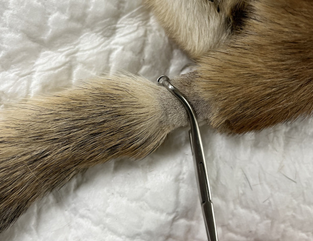
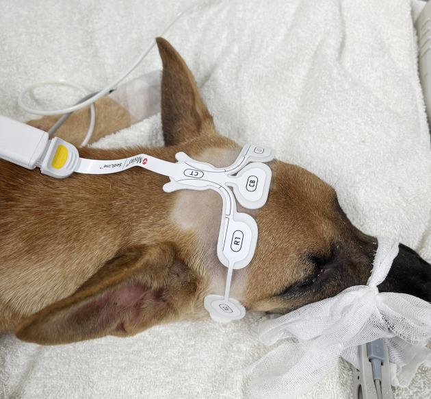
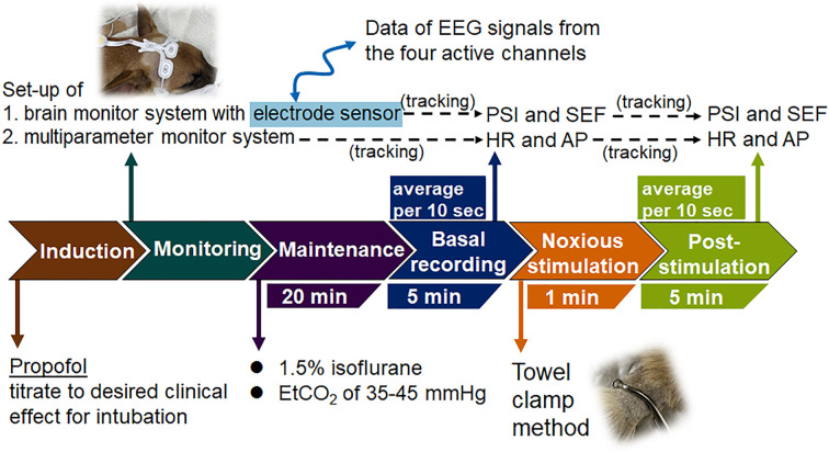
In veterinary clinical medicine, evaluating the balance between nociception and antinociception presents a great challenge for anesthesiologists during canine surgeries. Heart rate (HR) and mean arterial pressure (MAP) are suitable indexes for monitoring noxious stimuli during anesthesia. Frontal electroencephalography (EEG) records, including processed parameters, are recommended for evaluating nociceptive balance in anesthetized unconscious human patients, which is unexplored in veterinary medicine. Therefore, the objective is to explore the response of processed EEG parameters to noxious stimulation and elucidate the impact of noxious stimulation on frontal cortical activity in dogs anesthetized with 1.5% isoflurane. Fourteen dogs were included and underwent frontal EEG monitoring, measuring the patient state index (PSI) and spectral edge frequency (SEF) before and after administering noxious stimulation using the towel clamp method on the tail of each 1.5% isoflurane-anesthetized dog. As the noxious stimulation was applied, there was a simultaneous increase in PSI, HR, and MAP, with PSI exhibiting a drastic response. SEF, especially on the left side, also increased with noxious stimulation. In EEG power spectral analysis, the delta band was decreased, and the alpha and beta bands showed an increase following noxious stimulation, with a more profound elevation of beta band on the left side. This study suggests that noxious stimulation brings asymmetric frontal cortical arousal, changing brain activity by suppressing delta wave and augmenting alpha and beta waves. Consequently, PSI seems to be a potential indicator for detecting stimuli in canine isoflurane anesthesia.
期刊介绍:
The aim of this international journal is to accelerate progress in laboratory animal experimentation and disseminate relevant information in related areas through publication of peer reviewed Original papers and Review articles. The journal covers basic to applied biomedical research centering around use of experimental animals and also covers topics related to experimental animals such as technology, management, and animal welfare.


 分享
分享
 求助内容:
求助内容: 应助结果提醒方式:
 扫码关注我们
