{"title":"Pathological examination of factors involved in PD-L1 expression in patients with oral tongue squamous cell carcinoma.","authors":"Yu Koyama, Chiharu Ogawa, Chihiro Kurihara, Nao Hashimoto, Shota Shinagawa, Hiroya Okazaki, Takumi Koyama, Keisuke Sugahara, Akira Katakura","doi":"10.1186/s40902-024-00441-w","DOIUrl":null,"url":null,"abstract":"<p><strong>Background: </strong>Tumor tissues comprise cancer cells and stromal cells, and their interactions form the cancer microenvironment. Therefore, treatments targeting cells other than cancer cells are also actively being developed, and among them, treatment targeting PD-1, an immune checkpoint molecule that is important in tumor immune evasion, has also been indicated for head and neck cancer. PD-L1, a ligand of PD-1, is expressed in both tumor cells and stromal cells, and the scoring system based on the combined positivity rates of both types of cells, the combined positive score (CPS), is used for predicting treatment effect. However, much is unknown regarding the expression of PD-L1. In this study, we histopathologically examined factors controlling the expression of PD-1/PD-L1. This study included 37 patients who underwent resection surgery for tongue squamous cell carcinoma in the Department of Oral and Maxillofacial Surgery at Tokyo Dental College Suidobashi Hospital. The expression levels of PD-L1, α-SMA, and p53 were assessed by immunohistochemical staining.</p><p><strong>Results: </strong>Seven participants had CPS ≥ 20, twenty-four participants had 1 ≤ CPS < 20, and six participants had CPS < 1. The overall positivity rate of α-SMA, a marker for cancer-associated fibroblasts (CAFs), was 27% (10/37 participants), and the positivity rates of α-SMA for the three CPS groups were 85.7% (6/7 participants), 16.7% (4/24 participants), and 0% (0/6 participants), respectively. In addition, the overall positivity rate of p53 was 37.8% (14/37 participants), and the positivity rates of p53 for the three CPS groups were 71.4% (5/7 participants), 37.5% (9/24 participants), and 0% (0/6 participants), respectively.</p><p><strong>Conclusions: </strong>The expression of PD-L1 demonstrated an association with α-SMA and p53 positivity. In addition, compared with the expression of p53, the expression of α-SMA demonstrated a higher association with PD-L1 expression in patients with a high CPS. The abovementioned findings suggest that the interactions between CAFs, cancer cells, and immunocompetent cells may regulate the expression of PD-L1.</p>","PeriodicalId":18357,"journal":{"name":"Maxillofacial Plastic and Reconstructive Surgery","volume":"46 1","pages":"31"},"PeriodicalIF":2.8000,"publicationDate":"2024-08-08","publicationTypes":"Journal Article","fieldsOfStudy":null,"isOpenAccess":false,"openAccessPdf":"https://www.ncbi.nlm.nih.gov/pmc/articles/PMC11310371/pdf/","citationCount":"0","resultStr":null,"platform":"Semanticscholar","paperid":null,"PeriodicalName":"Maxillofacial Plastic and Reconstructive Surgery","FirstCategoryId":"1085","ListUrlMain":"https://doi.org/10.1186/s40902-024-00441-w","RegionNum":0,"RegionCategory":null,"ArticlePicture":[],"TitleCN":null,"AbstractTextCN":null,"PMCID":null,"EPubDate":"","PubModel":"","JCR":"Q2","JCRName":"DENTISTRY, ORAL SURGERY & MEDICINE","Score":null,"Total":0}
引用次数: 0
Abstract
Background: Tumor tissues comprise cancer cells and stromal cells, and their interactions form the cancer microenvironment. Therefore, treatments targeting cells other than cancer cells are also actively being developed, and among them, treatment targeting PD-1, an immune checkpoint molecule that is important in tumor immune evasion, has also been indicated for head and neck cancer. PD-L1, a ligand of PD-1, is expressed in both tumor cells and stromal cells, and the scoring system based on the combined positivity rates of both types of cells, the combined positive score (CPS), is used for predicting treatment effect. However, much is unknown regarding the expression of PD-L1. In this study, we histopathologically examined factors controlling the expression of PD-1/PD-L1. This study included 37 patients who underwent resection surgery for tongue squamous cell carcinoma in the Department of Oral and Maxillofacial Surgery at Tokyo Dental College Suidobashi Hospital. The expression levels of PD-L1, α-SMA, and p53 were assessed by immunohistochemical staining.
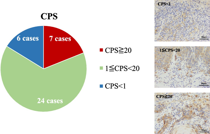
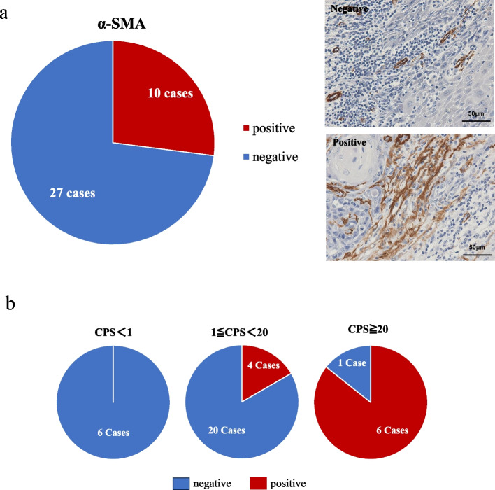
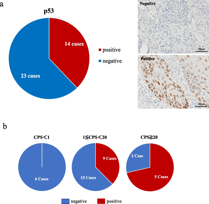
Results: Seven participants had CPS ≥ 20, twenty-four participants had 1 ≤ CPS < 20, and six participants had CPS < 1. The overall positivity rate of α-SMA, a marker for cancer-associated fibroblasts (CAFs), was 27% (10/37 participants), and the positivity rates of α-SMA for the three CPS groups were 85.7% (6/7 participants), 16.7% (4/24 participants), and 0% (0/6 participants), respectively. In addition, the overall positivity rate of p53 was 37.8% (14/37 participants), and the positivity rates of p53 for the three CPS groups were 71.4% (5/7 participants), 37.5% (9/24 participants), and 0% (0/6 participants), respectively.
Conclusions: The expression of PD-L1 demonstrated an association with α-SMA and p53 positivity. In addition, compared with the expression of p53, the expression of α-SMA demonstrated a higher association with PD-L1 expression in patients with a high CPS. The abovementioned findings suggest that the interactions between CAFs, cancer cells, and immunocompetent cells may regulate the expression of PD-L1.


 分享
分享
 求助内容:
求助内容: 应助结果提醒方式:
 扫码关注我们
