Utility of ER, p53, CEA and Napsin A in Histological Subtyping of Endometrial Carcinoma and Their Correlation with Clinicopathological Prognostic Parameters: Experience from a Referral Institute.
Saumya Shivakumar, Kausalya K Sahu, Ranjitha Rao, Chaithra Gv, Cheryl Sarah Philipose, Sharada Rai
{"title":"Utility of ER, p53, CEA and Napsin A in Histological Subtyping of Endometrial Carcinoma and Their Correlation with Clinicopathological Prognostic Parameters: Experience from a Referral Institute.","authors":"Saumya Shivakumar, Kausalya K Sahu, Ranjitha Rao, Chaithra Gv, Cheryl Sarah Philipose, Sharada Rai","doi":"10.30699/IJP.2024.2008693.3154","DOIUrl":null,"url":null,"abstract":"<p><strong>Background & objective: </strong>Endometrial Carcinoma (EC) is the most common gynecological cancer with a global incidence of 23.2 per 1 lakh population. Histological subclassification of EC is extremely crucial for the diagnosis, proper management strategies, and prognosis. This study was conducted in a tertiary care institute to analyze the expression pattern of a minimum panel of 4 markers (ER, p53, CEA, Napsin A) with emphasis on their utility in the routine histological subtyping, aberrant expression, and correlation with various clinicopathological parameters.</p><p><strong>Methods: </strong>A time-bound cross-sectional observational and analytical study was conducted, which includes cases diagnosed in our laboratory from January 2016 to April 2021.</p><p><strong>Results: </strong>Sixty cases diagnosed as EC during the study period formed the sample cases. The ER was expressed in 85% (53/60) of cases in the current study. Among them, 94% (50/53) were endometrioid endometrial carcinomas (EECs). A negative correlation was found between ER intensity and age (r= -1.48). Of 60 EC cases, 10 (16%) cases expressed p53. The tumors positive for p53 with higher intensity were negative for ER and vice versa. The expression pattern of ER and p53 was statistically significant (<i>P</i>=-0.021). On IHC, 84.6% (11/13) of CEA-positive cases expressed both ER and CEA, suggesting mucinous differentiation. Napsin A was expressed in two cases of EEC, FIGO grade I, and one case of serous carcinoma.</p><p><strong>Conclusion: </strong>An inverse association was found between ER and p53 expression. The CEA is valuable in identifying EEC with mucinous differentiation.</p>","PeriodicalId":38900,"journal":{"name":"Iranian Journal of Pathology","volume":"19 2","pages":"236-243"},"PeriodicalIF":0.0000,"publicationDate":"2024-01-01","publicationTypes":"Journal Article","fieldsOfStudy":null,"isOpenAccess":false,"openAccessPdf":"https://www.ncbi.nlm.nih.gov/pmc/articles/PMC11304467/pdf/","citationCount":"0","resultStr":null,"platform":"Semanticscholar","paperid":null,"PeriodicalName":"Iranian Journal of Pathology","FirstCategoryId":"1085","ListUrlMain":"https://doi.org/10.30699/IJP.2024.2008693.3154","RegionNum":0,"RegionCategory":null,"ArticlePicture":[],"TitleCN":null,"AbstractTextCN":null,"PMCID":null,"EPubDate":"2024/1/29 0:00:00","PubModel":"Epub","JCR":"Q3","JCRName":"Medicine","Score":null,"Total":0}
引用次数: 0
Abstract
Background & objective: Endometrial Carcinoma (EC) is the most common gynecological cancer with a global incidence of 23.2 per 1 lakh population. Histological subclassification of EC is extremely crucial for the diagnosis, proper management strategies, and prognosis. This study was conducted in a tertiary care institute to analyze the expression pattern of a minimum panel of 4 markers (ER, p53, CEA, Napsin A) with emphasis on their utility in the routine histological subtyping, aberrant expression, and correlation with various clinicopathological parameters.
Methods: A time-bound cross-sectional observational and analytical study was conducted, which includes cases diagnosed in our laboratory from January 2016 to April 2021.
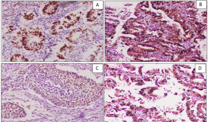
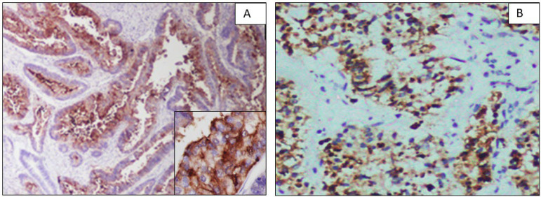
Results: Sixty cases diagnosed as EC during the study period formed the sample cases. The ER was expressed in 85% (53/60) of cases in the current study. Among them, 94% (50/53) were endometrioid endometrial carcinomas (EECs). A negative correlation was found between ER intensity and age (r= -1.48). Of 60 EC cases, 10 (16%) cases expressed p53. The tumors positive for p53 with higher intensity were negative for ER and vice versa. The expression pattern of ER and p53 was statistically significant (P=-0.021). On IHC, 84.6% (11/13) of CEA-positive cases expressed both ER and CEA, suggesting mucinous differentiation. Napsin A was expressed in two cases of EEC, FIGO grade I, and one case of serous carcinoma.
Conclusion: An inverse association was found between ER and p53 expression. The CEA is valuable in identifying EEC with mucinous differentiation.


 分享
分享
 求助内容:
求助内容: 应助结果提醒方式:
 扫码关注我们
