{"title":"A machine learning-based pipeline for multi-organ/tissue patient-specific radiation dosimetry in CT.","authors":"Eleftherios Tzanis, John Damilakis","doi":"10.1007/s00330-024-11002-0","DOIUrl":null,"url":null,"abstract":"<p><strong>Objectives: </strong>To develop a machine learning-based pipeline for multi-organ/tissue personalized radiation dosimetry in CT.</p><p><strong>Materials and methods: </strong>For the study, 95 chest CT scans and 85 abdominal CT scans were collected retrospectively. For each CT scan, a personalized Monte Carlo (MC) simulation was carried out. The produced 3D dose distributions and the respective CT examinations were utilized for the development of organ/tissue-specific dose prediction deep neural networks (DNNs). A pipeline that integrates a robust open-source organ segmentation tool with the dose prediction DNNs was developed for the automatic estimation of radiation doses for 30 organs/tissues including sub-volumes of the heart and lungs. The accuracy and time efficiency of the presented methodology was assessed. Statistical analysis (t-tests) was conducted to determine if the differences between the ground truth organ/tissue radiation dose estimates and the respective dose predictions were significant.</p><p><strong>Results: </strong>The lowest median percentage differences between MC-derived organ/tissue doses and DNN dose predictions were observed for the lung vessels (4.3%), small bowel (4.7%), pulmonary artery (4.7%), and colon (5.2%), while the highest differences were observed for the right lung's upper lobe (13.3%), spleen (13.1%), pancreas (12.1%), and stomach (11.6%). Statistical analysis showed that the differences were not significant (p-value > 0.18). Furthermore, the mean inference time, regarding the validation cohort, of the developed methodology was 77.0 ± 11.0 s.</p><p><strong>Conclusion: </strong>The proposed workflow enables fast and accurate organ/tissue radiation dose estimations. The developed algorithms and dose prediction DNNs are publicly available ( https://github.com/eltzanis/multi-structure-CT-dosimetry ).</p><p><strong>Clinical relevance statement: </strong>The accuracy and time efficiency of the developed pipeline compose a useful tool for personalized dosimetry in CT. By adopting the proposed workflow, institutions can utilize an automated pipeline for patient-specific dosimetry in CT.</p><p><strong>Key points: </strong>Personalized dosimetry is ideal, but is time-consuming. The proposed pipeline composes a tool for facilitating patient-specific CT dosimetry in routine clinical practice. The developed workflow integrates a robust open-source segmentation tool with organ/tissue-specific dose prediction neural networks.</p>","PeriodicalId":12076,"journal":{"name":"European Radiology","volume":" ","pages":"919-928"},"PeriodicalIF":4.7000,"publicationDate":"2025-02-01","publicationTypes":"Journal Article","fieldsOfStudy":null,"isOpenAccess":false,"openAccessPdf":"","citationCount":"0","resultStr":null,"platform":"Semanticscholar","paperid":null,"PeriodicalName":"European Radiology","FirstCategoryId":"3","ListUrlMain":"https://doi.org/10.1007/s00330-024-11002-0","RegionNum":2,"RegionCategory":"医学","ArticlePicture":[],"TitleCN":null,"AbstractTextCN":null,"PMCID":null,"EPubDate":"2024/8/13 0:00:00","PubModel":"Epub","JCR":"Q1","JCRName":"RADIOLOGY, NUCLEAR MEDICINE & MEDICAL IMAGING","Score":null,"Total":0}
引用次数: 0
Abstract
Objectives: To develop a machine learning-based pipeline for multi-organ/tissue personalized radiation dosimetry in CT.
Materials and methods: For the study, 95 chest CT scans and 85 abdominal CT scans were collected retrospectively. For each CT scan, a personalized Monte Carlo (MC) simulation was carried out. The produced 3D dose distributions and the respective CT examinations were utilized for the development of organ/tissue-specific dose prediction deep neural networks (DNNs). A pipeline that integrates a robust open-source organ segmentation tool with the dose prediction DNNs was developed for the automatic estimation of radiation doses for 30 organs/tissues including sub-volumes of the heart and lungs. The accuracy and time efficiency of the presented methodology was assessed. Statistical analysis (t-tests) was conducted to determine if the differences between the ground truth organ/tissue radiation dose estimates and the respective dose predictions were significant.
Results: The lowest median percentage differences between MC-derived organ/tissue doses and DNN dose predictions were observed for the lung vessels (4.3%), small bowel (4.7%), pulmonary artery (4.7%), and colon (5.2%), while the highest differences were observed for the right lung's upper lobe (13.3%), spleen (13.1%), pancreas (12.1%), and stomach (11.6%). Statistical analysis showed that the differences were not significant (p-value > 0.18). Furthermore, the mean inference time, regarding the validation cohort, of the developed methodology was 77.0 ± 11.0 s.
Conclusion: The proposed workflow enables fast and accurate organ/tissue radiation dose estimations. The developed algorithms and dose prediction DNNs are publicly available ( https://github.com/eltzanis/multi-structure-CT-dosimetry ).
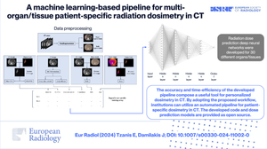
Clinical relevance statement: The accuracy and time efficiency of the developed pipeline compose a useful tool for personalized dosimetry in CT. By adopting the proposed workflow, institutions can utilize an automated pipeline for patient-specific dosimetry in CT.
Key points: Personalized dosimetry is ideal, but is time-consuming. The proposed pipeline composes a tool for facilitating patient-specific CT dosimetry in routine clinical practice. The developed workflow integrates a robust open-source segmentation tool with organ/tissue-specific dose prediction neural networks.
期刊介绍:
European Radiology (ER) continuously updates scientific knowledge in radiology by publication of strong original articles and state-of-the-art reviews written by leading radiologists. A well balanced combination of review articles, original papers, short communications from European radiological congresses and information on society matters makes ER an indispensable source for current information in this field.
This is the Journal of the European Society of Radiology, and the official journal of a number of societies.
From 2004-2008 supplements to European Radiology were published under its companion, European Radiology Supplements, ISSN 1613-3749.


 分享
分享
 求助内容:
求助内容: 应助结果提醒方式:
 扫码关注我们
