Rajeev A Essed, Yeva Prysiazhniuk, Ivar J Wamelink, Aynur Azizova, Vera C Keil
{"title":"Performance of amide proton transfer imaging to differentiate true progression from therapy-related changes in gliomas and metastases.","authors":"Rajeev A Essed, Yeva Prysiazhniuk, Ivar J Wamelink, Aynur Azizova, Vera C Keil","doi":"10.1007/s00330-024-11004-y","DOIUrl":null,"url":null,"abstract":"<p><strong>Objectives: </strong>Differentiating true progression or recurrence (TP/TR) from therapy-related changes (TRC) is complex in brain tumours. Amide proton transfer-weighted (APT) imaging is a chemical exchange saturation transfer (CEST) MRI technique that may improve diagnostic accuracy during radiological follow-up. This systematic review and meta-analysis elucidated the level of evidence and details of state-of-the-art imaging for APT-CEST in glioma and brain metastasis surveillance.</p><p><strong>Methods: </strong>PubMed, EMBASE, Web of Science, and Cochrane Library were systematically searched for original articles about glioma and metastasis patients who received APT-CEST imaging for suspected TP/TR within 2 years after (chemo)radiotherapy completion. Modified Quality Assessment of Diagnostic Accuracy Studies-2 criteria were applied. A meta-analysis was performed to pool results and to compare subgroups.</p><p><strong>Results: </strong>Fifteen studies were included for a narrative synthesis, twelve of which (500 patients) were deemed sufficiently homogeneous for a meta-analysis. Magnetisation transfer ratio asymmetry performed well in gliomas (sensitivity 0.88 [0.82-0.92], specificity 0.84 [0.72-0.91]) but not in metastases (sensitivity 0.64 [0.38-0.84], specificity 0.56 [0.33-0.77]). APT-CEST combined with conventional/advanced MRI rendered 0.92 [0.86-0.96] and 0.88 [0.72-0.95] in gliomas. Tumour type, TR prevalence, sex, and acquisition protocol were sources of significant inter-study heterogeneity in sensitivity (I<sup>2</sup> = 62.25%; p < 0.01) and specificity (I<sup>2</sup> = 66.31%; p < 0.001).</p><p><strong>Conclusion: </strong>A growing body of literature suggests that APT-CEST is a promising technique for improving the discrimination of TP/TR from TRC in gliomas, with limited data on metastases.</p><p><strong>Clinical relevance statement: </strong>This meta-analysis identified a utility for APT-CEST imaging regarding the non-invasive discrimination of brain tumour progression from therapy-related changes, providing a critical evaluation of sequence parameters and cut-off values, which can be used to improve response assessment and patient outcome.</p><p><strong>Key points: </strong>Therapy-related changes mimicking progression complicate brain tumour treatment. Amide proton imaging improves the non-invasive discrimination of glioma progression from therapy-related changes. Magnetisation transfer ratio asymmetry measurement seems not to have added value in brain metastases.</p>","PeriodicalId":12076,"journal":{"name":"European Radiology","volume":" ","pages":"580-591"},"PeriodicalIF":4.7000,"publicationDate":"2025-02-01","publicationTypes":"Journal Article","fieldsOfStudy":null,"isOpenAccess":false,"openAccessPdf":"https://www.ncbi.nlm.nih.gov/pmc/articles/PMC11782315/pdf/","citationCount":"0","resultStr":null,"platform":"Semanticscholar","paperid":null,"PeriodicalName":"European Radiology","FirstCategoryId":"3","ListUrlMain":"https://doi.org/10.1007/s00330-024-11004-y","RegionNum":2,"RegionCategory":"医学","ArticlePicture":[],"TitleCN":null,"AbstractTextCN":null,"PMCID":null,"EPubDate":"2024/8/12 0:00:00","PubModel":"Epub","JCR":"Q1","JCRName":"RADIOLOGY, NUCLEAR MEDICINE & MEDICAL IMAGING","Score":null,"Total":0}
引用次数: 0
Abstract
Objectives: Differentiating true progression or recurrence (TP/TR) from therapy-related changes (TRC) is complex in brain tumours. Amide proton transfer-weighted (APT) imaging is a chemical exchange saturation transfer (CEST) MRI technique that may improve diagnostic accuracy during radiological follow-up. This systematic review and meta-analysis elucidated the level of evidence and details of state-of-the-art imaging for APT-CEST in glioma and brain metastasis surveillance.
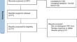
Methods: PubMed, EMBASE, Web of Science, and Cochrane Library were systematically searched for original articles about glioma and metastasis patients who received APT-CEST imaging for suspected TP/TR within 2 years after (chemo)radiotherapy completion. Modified Quality Assessment of Diagnostic Accuracy Studies-2 criteria were applied. A meta-analysis was performed to pool results and to compare subgroups.
Results: Fifteen studies were included for a narrative synthesis, twelve of which (500 patients) were deemed sufficiently homogeneous for a meta-analysis. Magnetisation transfer ratio asymmetry performed well in gliomas (sensitivity 0.88 [0.82-0.92], specificity 0.84 [0.72-0.91]) but not in metastases (sensitivity 0.64 [0.38-0.84], specificity 0.56 [0.33-0.77]). APT-CEST combined with conventional/advanced MRI rendered 0.92 [0.86-0.96] and 0.88 [0.72-0.95] in gliomas. Tumour type, TR prevalence, sex, and acquisition protocol were sources of significant inter-study heterogeneity in sensitivity (I2 = 62.25%; p < 0.01) and specificity (I2 = 66.31%; p < 0.001).
Conclusion: A growing body of literature suggests that APT-CEST is a promising technique for improving the discrimination of TP/TR from TRC in gliomas, with limited data on metastases.
Clinical relevance statement: This meta-analysis identified a utility for APT-CEST imaging regarding the non-invasive discrimination of brain tumour progression from therapy-related changes, providing a critical evaluation of sequence parameters and cut-off values, which can be used to improve response assessment and patient outcome.
Key points: Therapy-related changes mimicking progression complicate brain tumour treatment. Amide proton imaging improves the non-invasive discrimination of glioma progression from therapy-related changes. Magnetisation transfer ratio asymmetry measurement seems not to have added value in brain metastases.
期刊介绍:
European Radiology (ER) continuously updates scientific knowledge in radiology by publication of strong original articles and state-of-the-art reviews written by leading radiologists. A well balanced combination of review articles, original papers, short communications from European radiological congresses and information on society matters makes ER an indispensable source for current information in this field.
This is the Journal of the European Society of Radiology, and the official journal of a number of societies.
From 2004-2008 supplements to European Radiology were published under its companion, European Radiology Supplements, ISSN 1613-3749.


 分享
分享
 求助内容:
求助内容: 应助结果提醒方式:
 扫码关注我们
