{"title":"Simulation training in mammography with AI-generated images: a multireader study.","authors":"Krithika Rangarajan, Veeramakali Vignesh Manivannan, Harpinder Singh, Amit Gupta, Hrithik Maheshwari, Rishparn Gogoi, Debashish Gogoi, Rupam Jyoti Das, Smriti Hari, Surabhi Vyas, Raju Sharma, Shivam Pandey, V Seenu, Subhashis Banerjee, Vinay Namboodiri, Chetan Arora","doi":"10.1007/s00330-024-11005-x","DOIUrl":null,"url":null,"abstract":"<p><strong>Objectives: </strong>The interpretation of mammograms requires many years of training and experience. Currently, training in mammography, like the rest of diagnostic radiology, is through institutional libraries, books, and experience accumulated over time. We explore whether artificial Intelligence (AI)-generated images can help in simulation education and result in measurable improvement in performance of residents in training.</p><p><strong>Methods: </strong>We developed a generative adversarial network (GAN) that was capable of generating mammography images with varying characteristics, such as size and density, and created a tool with which a user could control these characteristics. The tool allowed the user (a radiology resident) to realistically insert cancers within different regions of the mammogram. We then provided this tool to residents in training. Residents were randomized into a practice group and a non-practice group, and the difference in performance before and after practice with such a tool (in comparison to no intervention in the non-practice group) was assessed.</p><p><strong>Results: </strong>Fifty residents participated in the study, 27 underwent simulation training, and 23 did not. There was a significant improvement in the sensitivity (7.43 percent, significant at p-value = 0.03), negative predictive value (5.05 percent, significant at p-value = 0.008) and accuracy (6.49 percent, significant at p-value = 0.01) among residents in the detection of cancer on mammograms after simulation training.</p><p><strong>Conclusion: </strong>Our study shows the value of simulation training in diagnostic radiology and explores the potential of generative AI to enable such simulation training.</p><p><strong>Clinical relevance statement: </strong>Using generative artificial intelligence, simulation training modules can be developed that can help residents in training by providing them with a visual impression of a variety of different cases.</p><p><strong>Key points: </strong>Generative networks can produce diagnostic imaging with specific characteristics, potentially useful for training residents. Training with generating images improved residents' mammographic diagnostic abilities. Development of a game-like interface that exploits these networks can result in improvement in performance over a short training period.</p>","PeriodicalId":12076,"journal":{"name":"European Radiology","volume":" ","pages":"562-571"},"PeriodicalIF":4.7000,"publicationDate":"2025-02-01","publicationTypes":"Journal Article","fieldsOfStudy":null,"isOpenAccess":false,"openAccessPdf":"","citationCount":"0","resultStr":null,"platform":"Semanticscholar","paperid":null,"PeriodicalName":"European Radiology","FirstCategoryId":"3","ListUrlMain":"https://doi.org/10.1007/s00330-024-11005-x","RegionNum":2,"RegionCategory":"医学","ArticlePicture":[],"TitleCN":null,"AbstractTextCN":null,"PMCID":null,"EPubDate":"2024/8/12 0:00:00","PubModel":"Epub","JCR":"Q1","JCRName":"RADIOLOGY, NUCLEAR MEDICINE & MEDICAL IMAGING","Score":null,"Total":0}
引用次数: 0
Abstract
Objectives: The interpretation of mammograms requires many years of training and experience. Currently, training in mammography, like the rest of diagnostic radiology, is through institutional libraries, books, and experience accumulated over time. We explore whether artificial Intelligence (AI)-generated images can help in simulation education and result in measurable improvement in performance of residents in training.
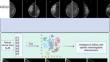
Methods: We developed a generative adversarial network (GAN) that was capable of generating mammography images with varying characteristics, such as size and density, and created a tool with which a user could control these characteristics. The tool allowed the user (a radiology resident) to realistically insert cancers within different regions of the mammogram. We then provided this tool to residents in training. Residents were randomized into a practice group and a non-practice group, and the difference in performance before and after practice with such a tool (in comparison to no intervention in the non-practice group) was assessed.
Results: Fifty residents participated in the study, 27 underwent simulation training, and 23 did not. There was a significant improvement in the sensitivity (7.43 percent, significant at p-value = 0.03), negative predictive value (5.05 percent, significant at p-value = 0.008) and accuracy (6.49 percent, significant at p-value = 0.01) among residents in the detection of cancer on mammograms after simulation training.
Conclusion: Our study shows the value of simulation training in diagnostic radiology and explores the potential of generative AI to enable such simulation training.
Clinical relevance statement: Using generative artificial intelligence, simulation training modules can be developed that can help residents in training by providing them with a visual impression of a variety of different cases.
Key points: Generative networks can produce diagnostic imaging with specific characteristics, potentially useful for training residents. Training with generating images improved residents' mammographic diagnostic abilities. Development of a game-like interface that exploits these networks can result in improvement in performance over a short training period.
目的:乳房 X 光检查的判读需要多年的培训和经验积累。目前,乳腺 X 线照相术的培训与其他放射诊断一样,都是通过机构图书馆、书籍和长期积累的经验进行的。我们探讨了人工智能(AI)生成的图像是否有助于模拟教学,并能显著提高住院医师在培训中的表现:我们开发了一个生成对抗网络(GAN),它能够生成具有不同特征(如大小和密度)的乳腺 X 射线图像,并创建了一个用户可以控制这些特征的工具。该工具允许用户(放射科住院医师)在乳房 X 光照片的不同区域真实地插入癌症。然后,我们将这一工具提供给正在接受培训的住院医师。住院医师被随机分为练习组和非练习组,并对使用该工具练习前后的表现差异(与非练习组的无干预相比)进行了评估:50 名住院医师参加了研究,其中 27 人接受了模拟训练,23 人未接受模拟训练。经过模拟训练后,住院医师在乳房 X 光检查中发现癌症的灵敏度(7.43%,p 值 = 0.03,有显著性意义)、阴性预测值(5.05%,p 值 = 0.008,有显著性意义)和准确度(6.49%,p 值 = 0.01,有显著性意义)均有明显提高:我们的研究显示了模拟训练在放射诊断学中的价值,并探索了生成式人工智能在实现此类模拟训练方面的潜力:利用生成式人工智能,可以开发出模拟训练模块,为住院医师提供各种不同病例的直观印象,从而帮助他们进行训练:关键点:生成网络可以生成具有特定特征的诊断图像,这对培训住院医师很有帮助。利用生成图像进行培训可提高住院医生的乳腺X线摄影诊断能力。利用这些网络开发类似游戏的界面可在短时间内提高培训效果。
期刊介绍:
European Radiology (ER) continuously updates scientific knowledge in radiology by publication of strong original articles and state-of-the-art reviews written by leading radiologists. A well balanced combination of review articles, original papers, short communications from European radiological congresses and information on society matters makes ER an indispensable source for current information in this field.
This is the Journal of the European Society of Radiology, and the official journal of a number of societies.
From 2004-2008 supplements to European Radiology were published under its companion, European Radiology Supplements, ISSN 1613-3749.


 分享
分享
 求助内容:
求助内容: 应助结果提醒方式:
 扫码关注我们
