{"title":"Thyroid hemiatrophy associated with papillary thyroid carcinoma.","authors":"Takuya Seko, Hiroki Kato, Tomohiro Ando, Kazuhiro Kobayashi, Hirofumi Shibata, Takenori Ogawa, Masaya Kawaguchi, Yoshifumi Noda, Fuminori Hyodo, Masayuki Matsuo","doi":"10.1007/s00234-024-03442-8","DOIUrl":null,"url":null,"abstract":"<p><strong>Purpose: </strong>The present study aimed to investigate CT imaging features, pathological findings, and prognosis in patients with thyroid hemiatrophy (THA) associated with papillary thyroid carcinoma (PTC).</p><p><strong>Methods: </strong>This retrospective study included 225 patients with histopathologically proven PTC treated by surgical resection who underwent preoperative CT scanning. On CT images, THA was defined as thyroid parenchymal hemiatrophy on the ipsilateral side of PTC. CT findings, overall survival, and disease-free survival were compared between patients with and without THA. Pathological findings were also assessed in PTCs with and without THA.</p><p><strong>Results: </strong>THA was observed in 35 of 225 (16%) patients with PTC. Atrophic thyroid parenchyma was observed in the right lobe of 20 patients (57%) and in the left lobe of the remaining 15 patients (43%). With respect to the solid components within PTCs, contrast-enhanced CT attenuation (114.2 ± 18.2 vs. 126.7 ± 31.3 HU; p < 0.05) and CT attenuation change for contrast-enhanced CT minus unenhanced CT (60.2 ± 18.1 vs. 72.3 ± 31.0 HU; p < 0.05) were significantly lower in PTCs with THA than in those without THA. Histopathologically, almost all PTCs with THA (97%) had keloid-like collagen, which is broad bundles of hypocellular collagen with bright eosinophilic hyalinization, typically observed in keloid. However, no significant differences were observed in the prognosis between the two groups.</p><p><strong>Conclusion: </strong>THA was occasionally observed in patients with PTC. Weak contrast-enhancement was distinct characteristic of PTC patients with THA, which is probably caused by keloid-like collagen.</p>","PeriodicalId":19422,"journal":{"name":"Neuroradiology","volume":" ","pages":"1795-1803"},"PeriodicalIF":2.6000,"publicationDate":"2024-10-01","publicationTypes":"Journal Article","fieldsOfStudy":null,"isOpenAccess":false,"openAccessPdf":"","citationCount":"0","resultStr":null,"platform":"Semanticscholar","paperid":null,"PeriodicalName":"Neuroradiology","FirstCategoryId":"3","ListUrlMain":"https://doi.org/10.1007/s00234-024-03442-8","RegionNum":3,"RegionCategory":"医学","ArticlePicture":[],"TitleCN":null,"AbstractTextCN":null,"PMCID":null,"EPubDate":"2024/8/13 0:00:00","PubModel":"Epub","JCR":"Q2","JCRName":"CLINICAL NEUROLOGY","Score":null,"Total":0}
引用次数: 0
Abstract
Purpose: The present study aimed to investigate CT imaging features, pathological findings, and prognosis in patients with thyroid hemiatrophy (THA) associated with papillary thyroid carcinoma (PTC).
Methods: This retrospective study included 225 patients with histopathologically proven PTC treated by surgical resection who underwent preoperative CT scanning. On CT images, THA was defined as thyroid parenchymal hemiatrophy on the ipsilateral side of PTC. CT findings, overall survival, and disease-free survival were compared between patients with and without THA. Pathological findings were also assessed in PTCs with and without THA.
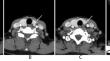
Results: THA was observed in 35 of 225 (16%) patients with PTC. Atrophic thyroid parenchyma was observed in the right lobe of 20 patients (57%) and in the left lobe of the remaining 15 patients (43%). With respect to the solid components within PTCs, contrast-enhanced CT attenuation (114.2 ± 18.2 vs. 126.7 ± 31.3 HU; p < 0.05) and CT attenuation change for contrast-enhanced CT minus unenhanced CT (60.2 ± 18.1 vs. 72.3 ± 31.0 HU; p < 0.05) were significantly lower in PTCs with THA than in those without THA. Histopathologically, almost all PTCs with THA (97%) had keloid-like collagen, which is broad bundles of hypocellular collagen with bright eosinophilic hyalinization, typically observed in keloid. However, no significant differences were observed in the prognosis between the two groups.
Conclusion: THA was occasionally observed in patients with PTC. Weak contrast-enhancement was distinct characteristic of PTC patients with THA, which is probably caused by keloid-like collagen.
期刊介绍:
Neuroradiology aims to provide state-of-the-art medical and scientific information in the fields of Neuroradiology, Neurosciences, Neurology, Psychiatry, Neurosurgery, and related medical specialities. Neuroradiology as the official Journal of the European Society of Neuroradiology receives submissions from all parts of the world and publishes peer-reviewed original research, comprehensive reviews, educational papers, opinion papers, and short reports on exceptional clinical observations and new technical developments in the field of Neuroimaging and Neurointervention. The journal has subsections for Diagnostic and Interventional Neuroradiology, Advanced Neuroimaging, Paediatric Neuroradiology, Head-Neck-ENT Radiology, Spine Neuroradiology, and for submissions from Japan. Neuroradiology aims to provide new knowledge about and insights into the function and pathology of the human nervous system that may help to better diagnose and treat nervous system diseases. Neuroradiology is a member of the Committee on Publication Ethics (COPE) and follows the COPE core practices. Neuroradiology prefers articles that are free of bias, self-critical regarding limitations, transparent and clear in describing study participants, methods, and statistics, and short in presenting results. Before peer-review all submissions are automatically checked by iThenticate to assess for potential overlap in prior publication.


 分享
分享
 求助内容:
求助内容: 应助结果提醒方式:
 扫码关注我们
