{"title":"Cervical internal carotid artery fenestration: a rare cause of lumen \"dissection''.","authors":"Natalia Valeria Pentara, Ioanna Koutroulou, Stephanos Finitsis, Vasileios Rafailidis, Elisavet Psoma, Nikolaos Grigoriadis, Panayiotis Prassopoulos, Theodoros Karapanayiotides","doi":"10.1007/s00276-024-03457-z","DOIUrl":null,"url":null,"abstract":"<p><strong>Purpose: </strong>To highlight the clinical and diagnostic importance of correctly identifying cervical internal carotid artery fenestration (fcICA), an extremely rare vascular anomaly, and to present a case where fcICA was initially misdiagnosed as a dissection in a patient with fibromuscular dysplasia (FMD).</p><p><strong>Methods: </strong>A 47-year-old woman with pulsatile tinnitus underwent computed tomography angiography (CTA) and digital subtraction angiography (DSA) to differentiate between fenestration and dissection of the internal carotid artery.</p><p><strong>Results: </strong>CTA revealed a fusiform dilatation of the distal C1 segment of the right internal carotid artery (ICA) with a linear filling defect, suggesting either fenestration or dissection. DSA confirmed the presence of a fenestrated right ICA segment composed of two symmetrical, smooth-walled limbs without a dissection flap, along with signs of FMD in the proximal vessel. The patient's symptoms were attributed to local flow perturbations induced by fcICA and FMD.</p><p><strong>Conclusion: </strong>This case illustrates that fcICA can be a true anatomical variant rather than a result of dissection, emphasizing the need for accurate imaging and diagnosis to avoid unnecessary treatments. The coexistence of fcICA with FMD increases the risk of dissection, necessitating careful monitoring. The distinction between fenestration and pseudofenestration remains challenging, requiring comprehensive imaging and close collaboration between radiologists and vascular neurologists.</p>","PeriodicalId":49461,"journal":{"name":"Surgical and Radiologic Anatomy","volume":" ","pages":"1659-1662"},"PeriodicalIF":1.2000,"publicationDate":"2024-10-01","publicationTypes":"Journal Article","fieldsOfStudy":null,"isOpenAccess":false,"openAccessPdf":"","citationCount":"0","resultStr":null,"platform":"Semanticscholar","paperid":null,"PeriodicalName":"Surgical and Radiologic Anatomy","FirstCategoryId":"3","ListUrlMain":"https://doi.org/10.1007/s00276-024-03457-z","RegionNum":4,"RegionCategory":"医学","ArticlePicture":[],"TitleCN":null,"AbstractTextCN":null,"PMCID":null,"EPubDate":"2024/8/13 0:00:00","PubModel":"Epub","JCR":"Q2","JCRName":"Medicine","Score":null,"Total":0}
引用次数: 0
Abstract
Purpose: To highlight the clinical and diagnostic importance of correctly identifying cervical internal carotid artery fenestration (fcICA), an extremely rare vascular anomaly, and to present a case where fcICA was initially misdiagnosed as a dissection in a patient with fibromuscular dysplasia (FMD).
Methods: A 47-year-old woman with pulsatile tinnitus underwent computed tomography angiography (CTA) and digital subtraction angiography (DSA) to differentiate between fenestration and dissection of the internal carotid artery.
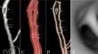
Results: CTA revealed a fusiform dilatation of the distal C1 segment of the right internal carotid artery (ICA) with a linear filling defect, suggesting either fenestration or dissection. DSA confirmed the presence of a fenestrated right ICA segment composed of two symmetrical, smooth-walled limbs without a dissection flap, along with signs of FMD in the proximal vessel. The patient's symptoms were attributed to local flow perturbations induced by fcICA and FMD.
Conclusion: This case illustrates that fcICA can be a true anatomical variant rather than a result of dissection, emphasizing the need for accurate imaging and diagnosis to avoid unnecessary treatments. The coexistence of fcICA with FMD increases the risk of dissection, necessitating careful monitoring. The distinction between fenestration and pseudofenestration remains challenging, requiring comprehensive imaging and close collaboration between radiologists and vascular neurologists.
期刊介绍:
Anatomy is a morphological science which cannot fail to interest the clinician. The practical application of anatomical research to clinical problems necessitates special adaptation and selectivity in choosing from numerous international works. Although there is a tendency to believe that meaningful advances in anatomy are unlikely, constant revision is necessary. Surgical and Radiologic Anatomy, the first international journal of Clinical anatomy has been created in this spirit.
Its goal is to serve clinicians, regardless of speciality-physicians, surgeons, radiologists or other specialists-as an indispensable aid with which they can improve their knowledge of anatomy. Each issue includes: Original papers, review articles, articles on the anatomical bases of medical, surgical and radiological techniques, articles of normal radiologic anatomy, brief reviews of anatomical publications of clinical interest.
Particular attention is given to high quality illustrations, which are indispensable for a better understanding of anatomical problems.
Surgical and Radiologic Anatomy is a journal written by anatomists for clinicians with a special interest in anatomy.


 分享
分享
 求助内容:
求助内容: 应助结果提醒方式:
 扫码关注我们
