Identifying the primary tumour in patients with cancer of unknown primary (CUP) using [18F]FDG PET/CT: a systematic review and individual patient data meta-analysis.
Jeroen R J Willemse, Doenja M J Lambregts, Sara Balduzzi, Winnie Schats, Petur Snaebjornsson, Serena Marchetti, Marieke A Vollebergh, Larissa W van Golen, Zing Cheung, Wouter V Vogel, Zuhir Bodalal, Sajjad Rostami, Oke Gerke, Tharani Sivakumaran, Regina G H Beets-Tan, Max J Lahaye
{"title":"Identifying the primary tumour in patients with cancer of unknown primary (CUP) using [<sup>18</sup>F]FDG PET/CT: a systematic review and individual patient data meta-analysis.","authors":"Jeroen R J Willemse, Doenja M J Lambregts, Sara Balduzzi, Winnie Schats, Petur Snaebjornsson, Serena Marchetti, Marieke A Vollebergh, Larissa W van Golen, Zing Cheung, Wouter V Vogel, Zuhir Bodalal, Sajjad Rostami, Oke Gerke, Tharani Sivakumaran, Regina G H Beets-Tan, Max J Lahaye","doi":"10.1007/s00259-024-06860-1","DOIUrl":null,"url":null,"abstract":"<p><strong>Purpose: </strong>In this systematic review and individual patient data (IPD) meta-analysis, we analysed the diagnostic performance of [<sup>18</sup>F]FDG PET/CT in detecting primary tumours in patients with CUP and evaluated whether the location of the predominant metastatic site influences the diagnostic performance.</p><p><strong>Methods: </strong>A systematic literature search from January 2005 to February 2024 was performed to identify articles describing the diagnostic performance of [<sup>18</sup>F]FDG PET/CT for primary tumour detection in CUP. Individual patient data retrieved from original articles or obtained from corresponding authors were grouped by the predominant metastatic site. The diagnostic performance of [<sup>18</sup>F]FDG PET/CT in detecting the underlying primary tumour was compared between predominant metastatic sites.</p><p><strong>Results: </strong>A total of 1865 patients from 32 studies were included. The largest subgroup included patients with predominant bone metastases (n = 622), followed by liver (n = 369), lymph node (n = 358), brain (n = 316), peritoneal (n = 70), lung (n = 67), and soft tissue (n = 23) metastases, leaving a small group of other/undefined metastases (n = 40). [<sup>18</sup>F]FDG PET/CT resulted in pooled detection rates to identify the primary tumour of 0.74 (for patients with predominant brain metastases), 0.54 (liver-predominant), 0.49 (bone-predominant), 0.46 (lung-predominant), 0.38 (peritoneal-predominant), 0.37 (lymph node-predominant), and 0.35 (soft-tissue-predominant).</p><p><strong>Conclusion: </strong>This individual patient data meta-analysis suggests that the ability of [<sup>18</sup>F]FDG PET/CT to identify the primary tumour in CUP depends on the distribution of metastatic sites. This finding emphasises the need for more tailored diagnostic approaches in different patient populations. In addition, alternative diagnostic tools, such as new PET tracers or whole-body (PET/)MRI, should be investigated.</p>","PeriodicalId":11909,"journal":{"name":"European Journal of Nuclear Medicine and Molecular Imaging","volume":" ","pages":"225-236"},"PeriodicalIF":8.6000,"publicationDate":"2024-12-01","publicationTypes":"Journal Article","fieldsOfStudy":null,"isOpenAccess":false,"openAccessPdf":"https://www.ncbi.nlm.nih.gov/pmc/articles/PMC11599304/pdf/","citationCount":"0","resultStr":null,"platform":"Semanticscholar","paperid":null,"PeriodicalName":"European Journal of Nuclear Medicine and Molecular Imaging","FirstCategoryId":"3","ListUrlMain":"https://doi.org/10.1007/s00259-024-06860-1","RegionNum":1,"RegionCategory":"医学","ArticlePicture":[],"TitleCN":null,"AbstractTextCN":null,"PMCID":null,"EPubDate":"2024/8/14 0:00:00","PubModel":"Epub","JCR":"Q1","JCRName":"RADIOLOGY, NUCLEAR MEDICINE & MEDICAL IMAGING","Score":null,"Total":0}
引用次数: 0
Abstract
Purpose: In this systematic review and individual patient data (IPD) meta-analysis, we analysed the diagnostic performance of [18F]FDG PET/CT in detecting primary tumours in patients with CUP and evaluated whether the location of the predominant metastatic site influences the diagnostic performance.
Methods: A systematic literature search from January 2005 to February 2024 was performed to identify articles describing the diagnostic performance of [18F]FDG PET/CT for primary tumour detection in CUP. Individual patient data retrieved from original articles or obtained from corresponding authors were grouped by the predominant metastatic site. The diagnostic performance of [18F]FDG PET/CT in detecting the underlying primary tumour was compared between predominant metastatic sites.
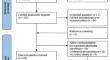
Results: A total of 1865 patients from 32 studies were included. The largest subgroup included patients with predominant bone metastases (n = 622), followed by liver (n = 369), lymph node (n = 358), brain (n = 316), peritoneal (n = 70), lung (n = 67), and soft tissue (n = 23) metastases, leaving a small group of other/undefined metastases (n = 40). [18F]FDG PET/CT resulted in pooled detection rates to identify the primary tumour of 0.74 (for patients with predominant brain metastases), 0.54 (liver-predominant), 0.49 (bone-predominant), 0.46 (lung-predominant), 0.38 (peritoneal-predominant), 0.37 (lymph node-predominant), and 0.35 (soft-tissue-predominant).
Conclusion: This individual patient data meta-analysis suggests that the ability of [18F]FDG PET/CT to identify the primary tumour in CUP depends on the distribution of metastatic sites. This finding emphasises the need for more tailored diagnostic approaches in different patient populations. In addition, alternative diagnostic tools, such as new PET tracers or whole-body (PET/)MRI, should be investigated.
期刊介绍:
The European Journal of Nuclear Medicine and Molecular Imaging serves as a platform for the exchange of clinical and scientific information within nuclear medicine and related professions. It welcomes international submissions from professionals involved in the functional, metabolic, and molecular investigation of diseases. The journal's coverage spans physics, dosimetry, radiation biology, radiochemistry, and pharmacy, providing high-quality peer review by experts in the field. Known for highly cited and downloaded articles, it ensures global visibility for research work and is part of the EJNMMI journal family.


 分享
分享

 求助内容:
求助内容: 应助结果提醒方式:
 扫码关注我们
