Daniel C Kargilis, Winnie Xu, Samir Reddy, Shilpa Shree Kuduva Ramesh, Steven Wang, Anh D Le, Chamith S Rajapakse
{"title":"Deep learning segmentation of mandible with lower dentition from cone beam CT.","authors":"Daniel C Kargilis, Winnie Xu, Samir Reddy, Shilpa Shree Kuduva Ramesh, Steven Wang, Anh D Le, Chamith S Rajapakse","doi":"10.1007/s11282-024-00770-6","DOIUrl":null,"url":null,"abstract":"<p><strong>Objectives: </strong>This study aimed to train a 3D U-Net convolutional neural network (CNN) for mandible and lower dentition segmentation from cone-beam computed tomography (CBCT) scans.</p><p><strong>Methods: </strong>In an ambispective cross-sectional design, CBCT scans from two hospitals (2009-2019 and 2021-2022) constituted an internal dataset and external validation set, respectively. Manual segmentation informed CNN training, and evaluations employed Dice similarity coefficient (DSC) for volumetric accuracy. A blinded oral maxillofacial surgeon performed qualitative grading of CBCT scans and object meshes. Statistical analyses included independent t-tests and ANOVA tests to compare DSC across patient subgroups of gender, race, body mass index (BMI), test dataset used, age, and degree of metal artifact. Tests were powered for a minimum detectable difference in DSC of 0.025, with alpha of 0.05 and power level of 0.8.</p><p><strong>Results: </strong>648 CBCT scans from 490 patients were included in the study. The CNN achieved high accuracy (average DSC: 0.945 internal, 0.940 external). No DSC differences were observed between test set used, gender, BMI, and race. Significant differences in DSC were identified based on age group and the degree of metal artifact. The majority (80%) of object meshes produced by both manual and automatic segmentation were rated as acceptable or higher quality.</p><p><strong>Conclusion: </strong>We developed a model for automatic mandible and lower dentition segmentation from CBCT scans in a demographically diverse cohort including a high degree of metal artifacts. The model demonstrated good accuracy on internal and external test sets, with majority acceptable quality from a clinical grader.</p>","PeriodicalId":56103,"journal":{"name":"Oral Radiology","volume":" ","pages":"1-9"},"PeriodicalIF":1.7000,"publicationDate":"2025-01-01","publicationTypes":"Journal Article","fieldsOfStudy":null,"isOpenAccess":false,"openAccessPdf":"","citationCount":"0","resultStr":null,"platform":"Semanticscholar","paperid":null,"PeriodicalName":"Oral Radiology","FirstCategoryId":"3","ListUrlMain":"https://doi.org/10.1007/s11282-024-00770-6","RegionNum":3,"RegionCategory":"医学","ArticlePicture":[],"TitleCN":null,"AbstractTextCN":null,"PMCID":null,"EPubDate":"2024/8/14 0:00:00","PubModel":"Epub","JCR":"Q3","JCRName":"DENTISTRY, ORAL SURGERY & MEDICINE","Score":null,"Total":0}
引用次数: 0
Abstract
Objectives: This study aimed to train a 3D U-Net convolutional neural network (CNN) for mandible and lower dentition segmentation from cone-beam computed tomography (CBCT) scans.
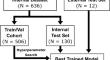
Methods: In an ambispective cross-sectional design, CBCT scans from two hospitals (2009-2019 and 2021-2022) constituted an internal dataset and external validation set, respectively. Manual segmentation informed CNN training, and evaluations employed Dice similarity coefficient (DSC) for volumetric accuracy. A blinded oral maxillofacial surgeon performed qualitative grading of CBCT scans and object meshes. Statistical analyses included independent t-tests and ANOVA tests to compare DSC across patient subgroups of gender, race, body mass index (BMI), test dataset used, age, and degree of metal artifact. Tests were powered for a minimum detectable difference in DSC of 0.025, with alpha of 0.05 and power level of 0.8.
Results: 648 CBCT scans from 490 patients were included in the study. The CNN achieved high accuracy (average DSC: 0.945 internal, 0.940 external). No DSC differences were observed between test set used, gender, BMI, and race. Significant differences in DSC were identified based on age group and the degree of metal artifact. The majority (80%) of object meshes produced by both manual and automatic segmentation were rated as acceptable or higher quality.
Conclusion: We developed a model for automatic mandible and lower dentition segmentation from CBCT scans in a demographically diverse cohort including a high degree of metal artifacts. The model demonstrated good accuracy on internal and external test sets, with majority acceptable quality from a clinical grader.
期刊介绍:
As the official English-language journal of the Japanese Society for Oral and Maxillofacial Radiology and the Asian Academy of Oral and Maxillofacial Radiology, Oral Radiology is intended to be a forum for international collaboration in head and neck diagnostic imaging and all related fields. Oral Radiology features cutting-edge research papers, review articles, case reports, and technical notes from both the clinical and experimental fields. As membership in the Society is not a prerequisite, contributions are welcome from researchers and clinicians worldwide.


 分享
分享
 求助内容:
求助内容: 应助结果提醒方式:
 扫码关注我们
