{"title":"Structural and functional characterization of archaeal DIMT1 unveils distinct protein dynamics essential for efficient catalysis","authors":"","doi":"10.1016/j.str.2024.07.013","DOIUrl":null,"url":null,"abstract":"<p>Dimethyladenosine transferase 1 (DIMT1), an ortholog of bacterial KsgA is a conserved protein that assists in ribosome biogenesis by modifying two successive adenosine bases near the 3′ end of small subunit (SSU) rRNA. Although KsgA/DIMT1 proteins have been characterized in bacteria and eukaryotes, they are yet unexplored in archaea. Also, their dynamics are not well understood. Here, we structurally and functionally characterized the apo and holo forms of archaeal DIMT1 from <em>Pyrococcus horikoshii</em>. Wild-type protein and mutants were analyzed to capture different transition states, including open, closed, and intermediate states. This study reports a unique inter-domain movement that is needed for substrate (RNA) positioning in the catalytic pocket, and is only observed in the presence of the cognate cofactors S-adenosyl-L-methionine (SAM) or S-adenosyl-L-homocysteine (SAH). The binding of the inhibitor sinefungine, an analog of SAM or SAH, to archaeal DIMT1 blocks the catalytic pocket and renders the enzyme inactive.</p>","PeriodicalId":22168,"journal":{"name":"Structure","volume":"77 1","pages":""},"PeriodicalIF":4.3000,"publicationDate":"2024-08-14","publicationTypes":"Journal Article","fieldsOfStudy":null,"isOpenAccess":false,"openAccessPdf":"","citationCount":"0","resultStr":null,"platform":"Semanticscholar","paperid":null,"PeriodicalName":"Structure","FirstCategoryId":"99","ListUrlMain":"https://doi.org/10.1016/j.str.2024.07.013","RegionNum":2,"RegionCategory":"生物学","ArticlePicture":[],"TitleCN":null,"AbstractTextCN":null,"PMCID":null,"EPubDate":"","PubModel":"","JCR":"Q2","JCRName":"BIOCHEMISTRY & MOLECULAR BIOLOGY","Score":null,"Total":0}
引用次数: 0
Abstract
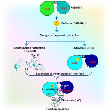
Dimethyladenosine transferase 1 (DIMT1), an ortholog of bacterial KsgA is a conserved protein that assists in ribosome biogenesis by modifying two successive adenosine bases near the 3′ end of small subunit (SSU) rRNA. Although KsgA/DIMT1 proteins have been characterized in bacteria and eukaryotes, they are yet unexplored in archaea. Also, their dynamics are not well understood. Here, we structurally and functionally characterized the apo and holo forms of archaeal DIMT1 from Pyrococcus horikoshii. Wild-type protein and mutants were analyzed to capture different transition states, including open, closed, and intermediate states. This study reports a unique inter-domain movement that is needed for substrate (RNA) positioning in the catalytic pocket, and is only observed in the presence of the cognate cofactors S-adenosyl-L-methionine (SAM) or S-adenosyl-L-homocysteine (SAH). The binding of the inhibitor sinefungine, an analog of SAM or SAH, to archaeal DIMT1 blocks the catalytic pocket and renders the enzyme inactive.
期刊介绍:
Structure aims to publish papers of exceptional interest in the field of structural biology. The journal strives to be essential reading for structural biologists, as well as biologists and biochemists that are interested in macromolecular structure and function. Structure strongly encourages the submission of manuscripts that present structural and molecular insights into biological function and mechanism. Other reports that address fundamental questions in structural biology, such as structure-based examinations of protein evolution, folding, and/or design, will also be considered. We will consider the application of any method, experimental or computational, at high or low resolution, to conduct structural investigations, as long as the method is appropriate for the biological, functional, and mechanistic question(s) being addressed. Likewise, reports describing single-molecule analysis of biological mechanisms are welcome.
In general, the editors encourage submission of experimental structural studies that are enriched by an analysis of structure-activity relationships and will not consider studies that solely report structural information unless the structure or analysis is of exceptional and broad interest. Studies reporting only homology models, de novo models, or molecular dynamics simulations are also discouraged unless the models are informed by or validated by novel experimental data; rationalization of a large body of existing experimental evidence and making testable predictions based on a model or simulation is often not considered sufficient.


 分享
分享
 求助内容:
求助内容: 应助结果提醒方式:
 扫码关注我们
