Multimodal imaging for the differential diagnosis and efficacy evaluation of intraocular retinoblastoma in children with selective ophthalmic artery infusion.
Jianshe Zhao, Ruodi Cui, Lin Li, Bing Zhao, Long Chen
{"title":"Multimodal imaging for the differential diagnosis and efficacy evaluation of intraocular retinoblastoma in children with selective ophthalmic artery infusion.","authors":"Jianshe Zhao, Ruodi Cui, Lin Li, Bing Zhao, Long Chen","doi":"10.21037/tp-24-2","DOIUrl":null,"url":null,"abstract":"<p><strong>Background: </strong>Retinoblastoma (RB) is the most common malignant tumor in children under the age of 3 years and is associated with a high disability and mortality rate. The aim of this study was, first, to evaluate the clinical efficacy of multimodal imaging in differentially diagnosing RB in children and in predicting the efficacy of selective ophthalmic artery infusion (SOAI) and, second, to identify the factors associated with this efficacy.</p><p><strong>Methods: </strong>This study retrospectively collected the data from 256 children with unilateral RB and intraocular involvement, including multimodal imaging magnetic resonance imaging (MRI), computed tomography (CT), and clinical characteristics. Among the cases, 33 with both CT and MRI data available were used to evaluate the diagnostic accuracy in distinguishing RB, with histopathological results serving as the gold standard. Additionally, a retrospective analysis was conducted on the MRI and clinical characteristics of 256 cases of unilateral RB with intraocular involvement before SOAI treatment. The predictive ability of imaging features and clinical characteristics for the treatment efficacy of children was analyzed, and the differences in globe salvage rates and visual preservation based on different tumor stages were evaluated.</p><p><strong>Results: </strong>The diagnostic accuracy of CT imaging for RB was 96.96% while that of MRI was 84.84%, with both showing high consistency with the histopathological results. CT images demonstrated a posterior intraocular mass with a high-density appearance, with spots, patches, or clustered calcifications visible within the tumor. The CT values were mostly above 100 Hounsfield units (HU), and enhanced scanning showed varying degrees of enhancement in noncalcified masses. MRI showed low or moderate signal intensity on T1-weighted images and moderate-to-high signal intensity on T2-weighted images, with significant enhancement after contrast administration. Tumors with more calcifications showed long T1 and short T2 signals. Patients with better prognosis had a higher delta signal increase (ΔSI), a greater distance from the optic disc, smaller tumor diameter, absence of implantation nodules or smaller implantation range, endogenous growth pattern, smaller extent of retinal detachment, absence of clinical high-risk factors, no vitreous hemorrhage, no globe shrinkage, and smaller calcification volume. The distance between the tumor and optic disc, clinical high-risk factors, and tumor growth pattern were found to be independent factors associated with prognosis. The rate of successful globe salvage and visual acuity decreased with increasing tumor stage.</p><p><strong>Conclusions: </strong>CT and MRI are highly valuable for the comprehensive assessment of tumors in pediatric RB. MRI alone can complete a comprehensive assessment of patients with RB and thus allow for the reduction radiation dose in children. Calcification of the tumor is crucial for diagnosis, and imaging findings can serve to inform patient prognosis and treatment planning. The distance between the tumor and optic disc, clinical high-risk factors, and tumor growth pattern are closely related to the prognosis of children.</p>","PeriodicalId":23294,"journal":{"name":"Translational pediatrics","volume":"13 7","pages":"1022-1032"},"PeriodicalIF":1.7000,"publicationDate":"2024-07-31","publicationTypes":"Journal Article","fieldsOfStudy":null,"isOpenAccess":false,"openAccessPdf":"https://www.ncbi.nlm.nih.gov/pmc/articles/PMC11320019/pdf/","citationCount":"0","resultStr":null,"platform":"Semanticscholar","paperid":null,"PeriodicalName":"Translational pediatrics","FirstCategoryId":"3","ListUrlMain":"https://doi.org/10.21037/tp-24-2","RegionNum":4,"RegionCategory":"医学","ArticlePicture":[],"TitleCN":null,"AbstractTextCN":null,"PMCID":null,"EPubDate":"2024/7/11 0:00:00","PubModel":"Epub","JCR":"Q2","JCRName":"PEDIATRICS","Score":null,"Total":0}
引用次数: 0
Abstract
Background: Retinoblastoma (RB) is the most common malignant tumor in children under the age of 3 years and is associated with a high disability and mortality rate. The aim of this study was, first, to evaluate the clinical efficacy of multimodal imaging in differentially diagnosing RB in children and in predicting the efficacy of selective ophthalmic artery infusion (SOAI) and, second, to identify the factors associated with this efficacy.
Methods: This study retrospectively collected the data from 256 children with unilateral RB and intraocular involvement, including multimodal imaging magnetic resonance imaging (MRI), computed tomography (CT), and clinical characteristics. Among the cases, 33 with both CT and MRI data available were used to evaluate the diagnostic accuracy in distinguishing RB, with histopathological results serving as the gold standard. Additionally, a retrospective analysis was conducted on the MRI and clinical characteristics of 256 cases of unilateral RB with intraocular involvement before SOAI treatment. The predictive ability of imaging features and clinical characteristics for the treatment efficacy of children was analyzed, and the differences in globe salvage rates and visual preservation based on different tumor stages were evaluated.
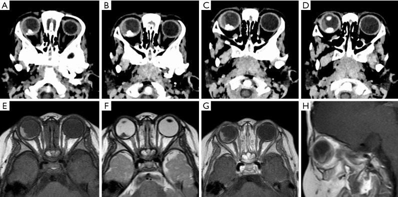
Results: The diagnostic accuracy of CT imaging for RB was 96.96% while that of MRI was 84.84%, with both showing high consistency with the histopathological results. CT images demonstrated a posterior intraocular mass with a high-density appearance, with spots, patches, or clustered calcifications visible within the tumor. The CT values were mostly above 100 Hounsfield units (HU), and enhanced scanning showed varying degrees of enhancement in noncalcified masses. MRI showed low or moderate signal intensity on T1-weighted images and moderate-to-high signal intensity on T2-weighted images, with significant enhancement after contrast administration. Tumors with more calcifications showed long T1 and short T2 signals. Patients with better prognosis had a higher delta signal increase (ΔSI), a greater distance from the optic disc, smaller tumor diameter, absence of implantation nodules or smaller implantation range, endogenous growth pattern, smaller extent of retinal detachment, absence of clinical high-risk factors, no vitreous hemorrhage, no globe shrinkage, and smaller calcification volume. The distance between the tumor and optic disc, clinical high-risk factors, and tumor growth pattern were found to be independent factors associated with prognosis. The rate of successful globe salvage and visual acuity decreased with increasing tumor stage.
Conclusions: CT and MRI are highly valuable for the comprehensive assessment of tumors in pediatric RB. MRI alone can complete a comprehensive assessment of patients with RB and thus allow for the reduction radiation dose in children. Calcification of the tumor is crucial for diagnosis, and imaging findings can serve to inform patient prognosis and treatment planning. The distance between the tumor and optic disc, clinical high-risk factors, and tumor growth pattern are closely related to the prognosis of children.


 分享
分享
 求助内容:
求助内容: 应助结果提醒方式:
 扫码关注我们
