{"title":"Effect of deep learning reconstruction on the assessment of pancreatic cystic lesions using computed tomography.","authors":"Jun Kanzawa, Koichiro Yasaka, Yuji Ohizumi, Yuichi Morita, Mariko Kurokawa, Osamu Abe","doi":"10.1007/s12194-024-00834-6","DOIUrl":null,"url":null,"abstract":"<p><p>This study aimed to compare the image quality and detection performance of pancreatic cystic lesions between computed tomography (CT) images reconstructed by deep learning reconstruction (DLR) and filtered back projection (FBP). This retrospective study included 54 patients (mean age: 67.7 ± 13.1) who underwent contrast-enhanced CT from May 2023 to August 2023. Among eligible patients, 30 and 24 were positive and negative for pancreatic cystic lesions, respectively. DLR and FBP were used to reconstruct portal venous phase images. Objective image quality analyses calculated quantitative image noise, signal-to-noise ratio (SNR), and contrast-to-noise ratio (CNR) using regions of interest on the abdominal aorta, pancreatic lesion, and pancreatic parenchyma. Three blinded radiologists performed subjective image quality assessment and lesion detection tests. Lesion depiction, normal structure illustration, subjective image noise, and overall image quality were utilized as subjective image quality indicators. DLR significantly reduced quantitative image noise compared with FBP (p < 0.001). SNR and CNR were significantly improved in DLR compared with FBP (p < 0.001). Three radiologists rated significantly higher scores for DLR in all subjective image quality indicators (p ≤ 0.029). Performance of DLR and FBP were comparable in lesion detection, with no statistically significant differences in the area under the receiver operating characteristic curve, sensitivity, specificity and accuracy. DLR reduced image noise and improved image quality with a clearer depiction of pancreatic structures. These improvements may have a positive effect on evaluating pancreatic cystic lesions, which can contribute to appropriate management of these lesions.</p>","PeriodicalId":46252,"journal":{"name":"Radiological Physics and Technology","volume":" ","pages":"827-833"},"PeriodicalIF":1.5000,"publicationDate":"2024-12-01","publicationTypes":"Journal Article","fieldsOfStudy":null,"isOpenAccess":false,"openAccessPdf":"https://www.ncbi.nlm.nih.gov/pmc/articles/PMC11579065/pdf/","citationCount":"0","resultStr":null,"platform":"Semanticscholar","paperid":null,"PeriodicalName":"Radiological Physics and Technology","FirstCategoryId":"1085","ListUrlMain":"https://doi.org/10.1007/s12194-024-00834-6","RegionNum":0,"RegionCategory":null,"ArticlePicture":[],"TitleCN":null,"AbstractTextCN":null,"PMCID":null,"EPubDate":"2024/8/15 0:00:00","PubModel":"Epub","JCR":"Q3","JCRName":"RADIOLOGY, NUCLEAR MEDICINE & MEDICAL IMAGING","Score":null,"Total":0}
引用次数: 0
Abstract
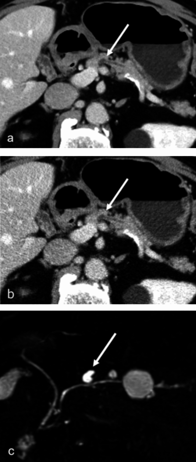
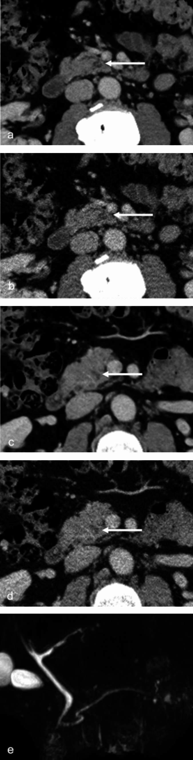
This study aimed to compare the image quality and detection performance of pancreatic cystic lesions between computed tomography (CT) images reconstructed by deep learning reconstruction (DLR) and filtered back projection (FBP). This retrospective study included 54 patients (mean age: 67.7 ± 13.1) who underwent contrast-enhanced CT from May 2023 to August 2023. Among eligible patients, 30 and 24 were positive and negative for pancreatic cystic lesions, respectively. DLR and FBP were used to reconstruct portal venous phase images. Objective image quality analyses calculated quantitative image noise, signal-to-noise ratio (SNR), and contrast-to-noise ratio (CNR) using regions of interest on the abdominal aorta, pancreatic lesion, and pancreatic parenchyma. Three blinded radiologists performed subjective image quality assessment and lesion detection tests. Lesion depiction, normal structure illustration, subjective image noise, and overall image quality were utilized as subjective image quality indicators. DLR significantly reduced quantitative image noise compared with FBP (p < 0.001). SNR and CNR were significantly improved in DLR compared with FBP (p < 0.001). Three radiologists rated significantly higher scores for DLR in all subjective image quality indicators (p ≤ 0.029). Performance of DLR and FBP were comparable in lesion detection, with no statistically significant differences in the area under the receiver operating characteristic curve, sensitivity, specificity and accuracy. DLR reduced image noise and improved image quality with a clearer depiction of pancreatic structures. These improvements may have a positive effect on evaluating pancreatic cystic lesions, which can contribute to appropriate management of these lesions.
期刊介绍:
The purpose of the journal Radiological Physics and Technology is to provide a forum for sharing new knowledge related to research and development in radiological science and technology, including medical physics and radiological technology in diagnostic radiology, nuclear medicine, and radiation therapy among many other radiological disciplines, as well as to contribute to progress and improvement in medical practice and patient health care.


 分享
分享
 求助内容:
求助内容: 应助结果提醒方式:
 扫码关注我们
