{"title":"Spatial arrangement, polarity, and posttranslational modifications of the microtubule system in the Drosophila eye.","authors":"Piotr Kos, Otto Baumann","doi":"10.1007/s00441-024-03914-6","DOIUrl":null,"url":null,"abstract":"<p><p>We have analyzed the organization of the microtubule system in photoreceptor cells and pigment cells within the adult Drosophila compound eye. Immunofluorescence localization of tubulin and of Short stop, a spectraplakin that has been reported to be involved in the anchorage of microtubule minus ends at the membrane, suggests the presence of non-centrosomal microtubule-organizing centers at the distal tip of the visual cells. Ultrastructural analyses confirm that microtubules emanate from membrane-associated plaques at the site of contact with cone cells and that all microtubules are aligned in distal-proximal direction within the photoreceptor cells. Determination of microtubule polarities demonstrated that about 95% of the microtubules in photoreceptor cells are oriented with their plus end in the direction of the synapse. Pigment cells in the eye contain only microtubules aligned in distal-proximal direction, with their plus end pointing towards the retinal floor. There, two populations of microtubules can be distinguished, single microtubules and bundled microtubules, the latter associated with actin filaments. Whereas microtubules in both photoreceptor cells and pigment cells are acetylated and mono/bi-glutamylated on α-tubulin, bundled microtubules in pigment cells are apparently also mono/bi-glutamylated on β-tubulin, providing the possibility of binding different microtubule-associated proteins.</p>","PeriodicalId":9712,"journal":{"name":"Cell and Tissue Research","volume":" ","pages":"123-137"},"PeriodicalIF":2.9000,"publicationDate":"2024-11-01","publicationTypes":"Journal Article","fieldsOfStudy":null,"isOpenAccess":false,"openAccessPdf":"https://www.ncbi.nlm.nih.gov/pmc/articles/PMC11525301/pdf/","citationCount":"0","resultStr":null,"platform":"Semanticscholar","paperid":null,"PeriodicalName":"Cell and Tissue Research","FirstCategoryId":"99","ListUrlMain":"https://doi.org/10.1007/s00441-024-03914-6","RegionNum":3,"RegionCategory":"生物学","ArticlePicture":[],"TitleCN":null,"AbstractTextCN":null,"PMCID":null,"EPubDate":"2024/8/17 0:00:00","PubModel":"Epub","JCR":"Q3","JCRName":"CELL BIOLOGY","Score":null,"Total":0}
引用次数: 0
Abstract
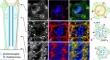
We have analyzed the organization of the microtubule system in photoreceptor cells and pigment cells within the adult Drosophila compound eye. Immunofluorescence localization of tubulin and of Short stop, a spectraplakin that has been reported to be involved in the anchorage of microtubule minus ends at the membrane, suggests the presence of non-centrosomal microtubule-organizing centers at the distal tip of the visual cells. Ultrastructural analyses confirm that microtubules emanate from membrane-associated plaques at the site of contact with cone cells and that all microtubules are aligned in distal-proximal direction within the photoreceptor cells. Determination of microtubule polarities demonstrated that about 95% of the microtubules in photoreceptor cells are oriented with their plus end in the direction of the synapse. Pigment cells in the eye contain only microtubules aligned in distal-proximal direction, with their plus end pointing towards the retinal floor. There, two populations of microtubules can be distinguished, single microtubules and bundled microtubules, the latter associated with actin filaments. Whereas microtubules in both photoreceptor cells and pigment cells are acetylated and mono/bi-glutamylated on α-tubulin, bundled microtubules in pigment cells are apparently also mono/bi-glutamylated on β-tubulin, providing the possibility of binding different microtubule-associated proteins.
期刊介绍:
The journal publishes regular articles and reviews in the areas of molecular, cell, and supracellular biology. In particular, the journal intends to provide a forum for publishing data that analyze the supracellular, integrative actions of gene products and their impact on the formation of tissue structure and function. Submission of papers with an emphasis on structure-function relationships as revealed by recombinant molecular technologies is especially encouraged. Areas of research with a long-standing tradition of publishing in Cell & Tissue Research include:
- neurobiology
- neuroendocrinology
- endocrinology
- reproductive biology
- skeletal and immune systems
- development
- stem cells
- muscle biology.


 分享
分享
 求助内容:
求助内容: 应助结果提醒方式:
 扫码关注我们
