R van den Elshout, B Ariëns, M Esmaeili, B Akkurt, M Mannil, F J A Meijer, A G van der Kolk, T W J Scheenen, D Henssen
{"title":"Distinguishing glioblastoma progression from treatment-related changes using DTI directionality growth analysis.","authors":"R van den Elshout, B Ariëns, M Esmaeili, B Akkurt, M Mannil, F J A Meijer, A G van der Kolk, T W J Scheenen, D Henssen","doi":"10.1007/s00234-024-03450-8","DOIUrl":null,"url":null,"abstract":"<p><strong>Background: </strong>It is difficult to distinguish between tumor progression (TP) and treatment-related abnormalities (TRA) in treated glioblastoma patients via conventional MRI, but this distinction is crucial for treatment decision making. Glioblastoma is known to exhibit an invasive growth pattern along white matter architecture and vasculature. This study quantified lesion development patterns in treated glioblastoma lesions and their relation to white matter microstructure to distinguish TP from TRA.</p><p><strong>Materials and methods: </strong>Glioblastoma patients with confirmed TP or TRA with T1-weighted contrast-enhanced and DTI MR scans from two posttreatment follow-up timepoints were reviewed. The contrast-enhancing regions were segmented, and the regions were coregistered to the DTI data. Lesion increase vectors were categorized into two groups: parallel (0-20 degrees) and perpendicular (70-90 degrees) to white matter. FA-values were also extracted. To test for a statistically significant difference between the TP and TRA groups, a Mann‒Whitney U test was performed.</p><p><strong>Results: </strong>Of 73 glioblastoma patients, fifteen were diagnosed with TRA, whereas 58 patients suffered TP. TP had a 25.8% (95% CI 24.1%-27.6%) increase in parallel lesions, and TRA had a 25.4% (95% CI 20.9%-29.9%) increase in parallel lesions. The perpendicular increase was 14.7% for TP (95% CI 13.0%-16.4%) and 18.0% (95% CI 13.5%-22.5%) for TRA. These results were not significantly different (p = 0.978). FA value for TP showed to be 0.248 (SD = 0.054) and for TRA it was 0.231 (SD = 0.075), showing no statistically significant difference (p = 0.121).</p><p><strong>Conclusions: </strong>Based on our results, quantifying posttreatment contrast-enhancing lesion development directionality with DTI in glioblastoma patients does not appear to effectively distinguish between TP and TRA.</p>","PeriodicalId":19422,"journal":{"name":"Neuroradiology","volume":" ","pages":"2143-2151"},"PeriodicalIF":2.6000,"publicationDate":"2024-12-01","publicationTypes":"Journal Article","fieldsOfStudy":null,"isOpenAccess":false,"openAccessPdf":"https://www.ncbi.nlm.nih.gov/pmc/articles/PMC11611950/pdf/","citationCount":"0","resultStr":null,"platform":"Semanticscholar","paperid":null,"PeriodicalName":"Neuroradiology","FirstCategoryId":"3","ListUrlMain":"https://doi.org/10.1007/s00234-024-03450-8","RegionNum":3,"RegionCategory":"医学","ArticlePicture":[],"TitleCN":null,"AbstractTextCN":null,"PMCID":null,"EPubDate":"2024/8/17 0:00:00","PubModel":"Epub","JCR":"Q2","JCRName":"CLINICAL NEUROLOGY","Score":null,"Total":0}
引用次数: 0
Abstract
Background: It is difficult to distinguish between tumor progression (TP) and treatment-related abnormalities (TRA) in treated glioblastoma patients via conventional MRI, but this distinction is crucial for treatment decision making. Glioblastoma is known to exhibit an invasive growth pattern along white matter architecture and vasculature. This study quantified lesion development patterns in treated glioblastoma lesions and their relation to white matter microstructure to distinguish TP from TRA.
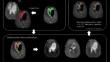
Materials and methods: Glioblastoma patients with confirmed TP or TRA with T1-weighted contrast-enhanced and DTI MR scans from two posttreatment follow-up timepoints were reviewed. The contrast-enhancing regions were segmented, and the regions were coregistered to the DTI data. Lesion increase vectors were categorized into two groups: parallel (0-20 degrees) and perpendicular (70-90 degrees) to white matter. FA-values were also extracted. To test for a statistically significant difference between the TP and TRA groups, a Mann‒Whitney U test was performed.
Results: Of 73 glioblastoma patients, fifteen were diagnosed with TRA, whereas 58 patients suffered TP. TP had a 25.8% (95% CI 24.1%-27.6%) increase in parallel lesions, and TRA had a 25.4% (95% CI 20.9%-29.9%) increase in parallel lesions. The perpendicular increase was 14.7% for TP (95% CI 13.0%-16.4%) and 18.0% (95% CI 13.5%-22.5%) for TRA. These results were not significantly different (p = 0.978). FA value for TP showed to be 0.248 (SD = 0.054) and for TRA it was 0.231 (SD = 0.075), showing no statistically significant difference (p = 0.121).
Conclusions: Based on our results, quantifying posttreatment contrast-enhancing lesion development directionality with DTI in glioblastoma patients does not appear to effectively distinguish between TP and TRA.
期刊介绍:
Neuroradiology aims to provide state-of-the-art medical and scientific information in the fields of Neuroradiology, Neurosciences, Neurology, Psychiatry, Neurosurgery, and related medical specialities. Neuroradiology as the official Journal of the European Society of Neuroradiology receives submissions from all parts of the world and publishes peer-reviewed original research, comprehensive reviews, educational papers, opinion papers, and short reports on exceptional clinical observations and new technical developments in the field of Neuroimaging and Neurointervention. The journal has subsections for Diagnostic and Interventional Neuroradiology, Advanced Neuroimaging, Paediatric Neuroradiology, Head-Neck-ENT Radiology, Spine Neuroradiology, and for submissions from Japan. Neuroradiology aims to provide new knowledge about and insights into the function and pathology of the human nervous system that may help to better diagnose and treat nervous system diseases. Neuroradiology is a member of the Committee on Publication Ethics (COPE) and follows the COPE core practices. Neuroradiology prefers articles that are free of bias, self-critical regarding limitations, transparent and clear in describing study participants, methods, and statistics, and short in presenting results. Before peer-review all submissions are automatically checked by iThenticate to assess for potential overlap in prior publication.


 分享
分享
 求助内容:
求助内容: 应助结果提醒方式:
 扫码关注我们
