Kristoffer Toldnes Cumming, Stefan Markus Reitzner, Marit Hanslien, Kenneth Skilnand, Olivier R. Seynnes, Oscar Horwath, Niklas Psilander, Carl Johan Sundberg, Truls Raastad
{"title":"Muscle memory in humans: evidence for myonuclear permanence and long-term transcriptional regulation after strength training","authors":"Kristoffer Toldnes Cumming, Stefan Markus Reitzner, Marit Hanslien, Kenneth Skilnand, Olivier R. Seynnes, Oscar Horwath, Niklas Psilander, Carl Johan Sundberg, Truls Raastad","doi":"10.1113/JP285675","DOIUrl":null,"url":null,"abstract":"<div>\n \n <section>\n \n \n <div>The objective of this work was to investigate myonuclear permanence and transcriptional regulation as mechanisms for cellular muscle memory after strength training in humans. Twelve untrained men and women performed 10 weeks of unilateral elbow-flexor strength training followed by 16 weeks of de-training. Thereafter, 10 weeks’ re-training was conducted with both arms: the previously trained arm and the contralateral untrained control arm. Muscle biopsies were taken from the trained arm before and after both training periods and from the control arm before and after re-training. Muscle biopsies were analysed for fibre cross-sectional area (fCSA), myonuclei and global transcriptomics (RNA sequencing). During the first training period, myonuclei increased in type 1 (13 ± 17%) and type 2 (33 ± 23%) fibres together with a 30 ± 43% non-significant increase in mixed fibre fCSA (<i>P</i> = 0.069). Following de-training, fCSA decreased in both fibre types, whereas myonuclei were maintained, resulting in 33% higher myonuclear number in previously trained <i>vs</i>. control muscle in type 2 fibres. Furthermore, in the previously trained muscle, three differentially expressed genes (DEGs; <i>EGR1</i>, <i>MYL5</i> and <i>COL1A1</i>) were observed. Following re-training, the previously trained muscle showed larger type 2 fCSA compared to the control (<i>P</i> = 0.035). However, delta change in type 2 fCSA was not different between muscles. Gene expression was more dramatically changed in the control arm (1338 DEGs) than in the previously trained arm (822 DEGs). The sustained higher number of myonuclei in the previously trained muscle confirms myonuclear accretion and permanence in humans. Nevertheless, because of the unclear effect on the subsequent hypertrophy with re-training, the physiological benefit remains to be determined.\n\n <figure>\n <div><picture>\n <source></source></picture><p></p>\n </div>\n </figure>\n </div>\n </section>\n \n <section>\n \n <h3> Key points</h3>\n \n <div>\n <ul>\n \n <li>Muscle memory is a cellular mechanism that describes the capacity of skeletal muscle fibres to respond differently to training stimuli if the stimuli have been previously encountered.</li>\n \n <li>This study overcomes past methodological limitations related to the choice of muscles and analytical procedures.</li>\n \n <li>We show that myonuclear number is increased after strength training and maintained during de-training.</li>\n \n <li>Increased myonuclear number and differentially expressed genes related to muscle performance and development in the previously trained muscle did not translate into a clearly superior responses during re-training. Because of the unclear effect on the subsequent hypertrophy and muscle strength gain with re-training, the physiological benefit remains to be determined.</li>\n </ul>\n </div>\n </section>\n </div>","PeriodicalId":50088,"journal":{"name":"Journal of Physiology-London","volume":"602 17","pages":"4171-4193"},"PeriodicalIF":4.4000,"publicationDate":"2024-08-19","publicationTypes":"Journal Article","fieldsOfStudy":null,"isOpenAccess":false,"openAccessPdf":"https://onlinelibrary.wiley.com/doi/epdf/10.1113/JP285675","citationCount":"0","resultStr":null,"platform":"Semanticscholar","paperid":null,"PeriodicalName":"Journal of Physiology-London","FirstCategoryId":"3","ListUrlMain":"https://physoc.onlinelibrary.wiley.com/doi/10.1113/JP285675","RegionNum":2,"RegionCategory":"医学","ArticlePicture":[],"TitleCN":null,"AbstractTextCN":null,"PMCID":null,"EPubDate":"","PubModel":"","JCR":"Q1","JCRName":"NEUROSCIENCES","Score":null,"Total":0}
引用次数: 0
Abstract
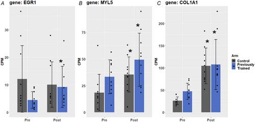
The objective of this work was to investigate myonuclear permanence and transcriptional regulation as mechanisms for cellular muscle memory after strength training in humans. Twelve untrained men and women performed 10 weeks of unilateral elbow-flexor strength training followed by 16 weeks of de-training. Thereafter, 10 weeks’ re-training was conducted with both arms: the previously trained arm and the contralateral untrained control arm. Muscle biopsies were taken from the trained arm before and after both training periods and from the control arm before and after re-training. Muscle biopsies were analysed for fibre cross-sectional area (fCSA), myonuclei and global transcriptomics (RNA sequencing). During the first training period, myonuclei increased in type 1 (13 ± 17%) and type 2 (33 ± 23%) fibres together with a 30 ± 43% non-significant increase in mixed fibre fCSA (P = 0.069). Following de-training, fCSA decreased in both fibre types, whereas myonuclei were maintained, resulting in 33% higher myonuclear number in previously trained vs. control muscle in type 2 fibres. Furthermore, in the previously trained muscle, three differentially expressed genes (DEGs; EGR1, MYL5 and COL1A1) were observed. Following re-training, the previously trained muscle showed larger type 2 fCSA compared to the control (P = 0.035). However, delta change in type 2 fCSA was not different between muscles. Gene expression was more dramatically changed in the control arm (1338 DEGs) than in the previously trained arm (822 DEGs). The sustained higher number of myonuclei in the previously trained muscle confirms myonuclear accretion and permanence in humans. Nevertheless, because of the unclear effect on the subsequent hypertrophy with re-training, the physiological benefit remains to be determined.
Key points
Muscle memory is a cellular mechanism that describes the capacity of skeletal muscle fibres to respond differently to training stimuli if the stimuli have been previously encountered.
This study overcomes past methodological limitations related to the choice of muscles and analytical procedures.
We show that myonuclear number is increased after strength training and maintained during de-training.
Increased myonuclear number and differentially expressed genes related to muscle performance and development in the previously trained muscle did not translate into a clearly superior responses during re-training. Because of the unclear effect on the subsequent hypertrophy and muscle strength gain with re-training, the physiological benefit remains to be determined.
期刊介绍:
The Journal of Physiology publishes full-length original Research Papers and Techniques for Physiology, which are short papers aimed at disseminating new techniques for physiological research. Articles solicited by the Editorial Board include Perspectives, Symposium Reports and Topical Reviews, which highlight areas of special physiological interest. CrossTalk articles are short editorial-style invited articles framing a debate between experts in the field on controversial topics. Letters to the Editor and Journal Club articles are also published. All categories of papers are subjected to peer reivew.
The Journal of Physiology welcomes submitted research papers in all areas of physiology. Authors should present original work that illustrates new physiological principles or mechanisms. Papers on work at the molecular level, at the level of the cell membrane, single cells, tissues or organs and on systems physiology are all acceptable. Theoretical papers and papers that use computational models to further our understanding of physiological processes will be considered if based on experimentally derived data and if the hypothesis advanced is directly amenable to experimental testing. While emphasis is on human and mammalian physiology, work on lower vertebrate or invertebrate preparations may be suitable if it furthers the understanding of the functioning of other organisms including mammals.


 分享
分享
 求助内容:
求助内容: 应助结果提醒方式:
 扫码关注我们
