Dowoon Han, Subin Kim, Jae-Hoon Jung, Ki-Hong Chang
{"title":"Cavernous Hemangioma of the Mastoid Antrum.","authors":"Dowoon Han, Subin Kim, Jae-Hoon Jung, Ki-Hong Chang","doi":"10.5152/iao.2024.231171","DOIUrl":null,"url":null,"abstract":"<p><p>Hemangioma is a common vascular neoplasm that arises in the head and neck regions but is rare in the petrous bone. We report the first case of a solitary cavernous hemangioma in the mastoid antrum. A 68-year-old woman visited our hospital with a complaint of tinnitus without any other symptoms. Tinnitus of the right ear occurred especially when the patient yawned or swallowed. Both tympanic membranes appeared normal on otoscopic examination. On pure-tone audiometry, mild hearing loss up to 25 dB was detected in the right ear. Temporal bone computed tomography revealed a 7.0 mm × 4.5 mm × 5 mm, solitary soft tissue mass in the aditus ad antrum. Excisional biopsy was performed under general anesthesia through the canal wall as in a mastoidectomy. The mass was completely removed without any bleeding or ossicular chain damage. The mass was confirmed as a cavernous hemangioma. During follow-up, the patient's tinnitus and right low-tone hearing loss improved. No solitary hemangioma of the mastoid antrum has been reported previously. Surgical excision of the lesion appears to be proper treatment to achieve pathologic confirmation along with resolution of symptoms.</p>","PeriodicalId":94238,"journal":{"name":"The journal of international advanced otology","volume":"20 4","pages":"372-374"},"PeriodicalIF":1.2000,"publicationDate":"2024-07-29","publicationTypes":"Journal Article","fieldsOfStudy":null,"isOpenAccess":false,"openAccessPdf":"https://www.ncbi.nlm.nih.gov/pmc/articles/PMC11363169/pdf/","citationCount":"0","resultStr":null,"platform":"Semanticscholar","paperid":null,"PeriodicalName":"The journal of international advanced otology","FirstCategoryId":"1085","ListUrlMain":"https://doi.org/10.5152/iao.2024.231171","RegionNum":0,"RegionCategory":null,"ArticlePicture":[],"TitleCN":null,"AbstractTextCN":null,"PMCID":null,"EPubDate":"","PubModel":"","JCR":"","JCRName":"","Score":null,"Total":0}
引用次数: 0
Abstract
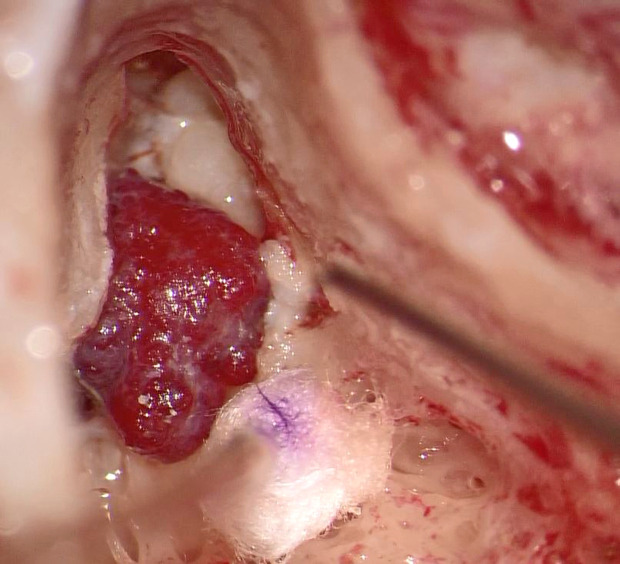
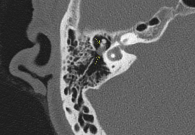
Hemangioma is a common vascular neoplasm that arises in the head and neck regions but is rare in the petrous bone. We report the first case of a solitary cavernous hemangioma in the mastoid antrum. A 68-year-old woman visited our hospital with a complaint of tinnitus without any other symptoms. Tinnitus of the right ear occurred especially when the patient yawned or swallowed. Both tympanic membranes appeared normal on otoscopic examination. On pure-tone audiometry, mild hearing loss up to 25 dB was detected in the right ear. Temporal bone computed tomography revealed a 7.0 mm × 4.5 mm × 5 mm, solitary soft tissue mass in the aditus ad antrum. Excisional biopsy was performed under general anesthesia through the canal wall as in a mastoidectomy. The mass was completely removed without any bleeding or ossicular chain damage. The mass was confirmed as a cavernous hemangioma. During follow-up, the patient's tinnitus and right low-tone hearing loss improved. No solitary hemangioma of the mastoid antrum has been reported previously. Surgical excision of the lesion appears to be proper treatment to achieve pathologic confirmation along with resolution of symptoms.


 分享
分享

 求助内容:
求助内容: 应助结果提醒方式:
 扫码关注我们
