Sam Van Roy, Liangge Hsu, Joseph Ho, Benjamin M Scirica, David Fischer, Samuel B Snider, Jong Woo Lee
{"title":"Quantitative and Radiological Assessment of Post-cardiac-Arrest Comatose Patients with Diffusion-Weighted Magnetic Resonance Imaging.","authors":"Sam Van Roy, Liangge Hsu, Joseph Ho, Benjamin M Scirica, David Fischer, Samuel B Snider, Jong Woo Lee","doi":"10.1007/s12028-024-02087-y","DOIUrl":null,"url":null,"abstract":"<p><strong>Background: </strong>Although magnetic resonance imaging, particularly diffusion-weighted imaging, has increasingly been used as part of a multimodal approach to prognostication in patients who are comatose after cardiac arrest, the performance of quantitative analysis of apparent diffusion coefficient (ADC) maps, as compared to standard radiologist impression, has not been well characterized. This retrospective study evaluated quantitative ADC analysis to the identification of anoxic brain injury by diffusion abnormalities on standard clinical magnetic resonance imaging reports.</p><p><strong>Methods: </strong>The cohort included 204 previously described comatose patients after cardiac arrest. Clinical outcome was assessed by (1) 3-6 month post-cardiac-arrest cerebral performance category and (2) coma recovery to following commands. Radiological evaluation was obtained from clinical reports and characterized as diffuse, cortex only, deep gray matter structures only, or no anoxic injury. Quantitative analyses of ADC maps were obtained in specific regions of interest (ROIs), whole cortex, and whole brain. A subgroup analysis of 172 was performed after eliminating images with artifacts and preexisting lesions.</p><p><strong>Results: </strong>Radiological assessment outperformed quantitative assessment over all evaluated regions (area under the curve [AUC] 0.80 for radiological interpretation and 0.70 for the occipital region, the best performing ROI, p = 0.011); agreement was substantial for all regions. Radiological assessment still outperformed quantitative analysis in the subgroup analysis, though by smaller margins and with substantial to near-perfect agreement. When assessing for coma recovery only, the difference was no longer significant (AUC 0.83 vs. 0.81 for the occipital region, p = 0.70).</p><p><strong>Conclusions: </strong>Although quantitative analysis eliminates interrater differences in the interpretation of abnormal diffusion imaging and avoids bias from other prediction modalities, clinical radiologist interpretation has a higher predictive value for outcome. Agreement between radiological and quantitative analysis improved when using high-quality scans and when assessing for coma recovery using following commands. Quantitative assessment may thus be more subject to variability in both clinical management and scan quality than radiological assessment.</p>","PeriodicalId":19118,"journal":{"name":"Neurocritical Care","volume":" ","pages":"541-550"},"PeriodicalIF":3.6000,"publicationDate":"2025-04-01","publicationTypes":"Journal Article","fieldsOfStudy":null,"isOpenAccess":false,"openAccessPdf":"","citationCount":"0","resultStr":null,"platform":"Semanticscholar","paperid":null,"PeriodicalName":"Neurocritical Care","FirstCategoryId":"3","ListUrlMain":"https://doi.org/10.1007/s12028-024-02087-y","RegionNum":3,"RegionCategory":"医学","ArticlePicture":[],"TitleCN":null,"AbstractTextCN":null,"PMCID":null,"EPubDate":"2024/8/20 0:00:00","PubModel":"Epub","JCR":"Q2","JCRName":"CLINICAL NEUROLOGY","Score":null,"Total":0}
引用次数: 0
Abstract
Background: Although magnetic resonance imaging, particularly diffusion-weighted imaging, has increasingly been used as part of a multimodal approach to prognostication in patients who are comatose after cardiac arrest, the performance of quantitative analysis of apparent diffusion coefficient (ADC) maps, as compared to standard radiologist impression, has not been well characterized. This retrospective study evaluated quantitative ADC analysis to the identification of anoxic brain injury by diffusion abnormalities on standard clinical magnetic resonance imaging reports.
Methods: The cohort included 204 previously described comatose patients after cardiac arrest. Clinical outcome was assessed by (1) 3-6 month post-cardiac-arrest cerebral performance category and (2) coma recovery to following commands. Radiological evaluation was obtained from clinical reports and characterized as diffuse, cortex only, deep gray matter structures only, or no anoxic injury. Quantitative analyses of ADC maps were obtained in specific regions of interest (ROIs), whole cortex, and whole brain. A subgroup analysis of 172 was performed after eliminating images with artifacts and preexisting lesions.
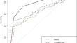
Results: Radiological assessment outperformed quantitative assessment over all evaluated regions (area under the curve [AUC] 0.80 for radiological interpretation and 0.70 for the occipital region, the best performing ROI, p = 0.011); agreement was substantial for all regions. Radiological assessment still outperformed quantitative analysis in the subgroup analysis, though by smaller margins and with substantial to near-perfect agreement. When assessing for coma recovery only, the difference was no longer significant (AUC 0.83 vs. 0.81 for the occipital region, p = 0.70).
Conclusions: Although quantitative analysis eliminates interrater differences in the interpretation of abnormal diffusion imaging and avoids bias from other prediction modalities, clinical radiologist interpretation has a higher predictive value for outcome. Agreement between radiological and quantitative analysis improved when using high-quality scans and when assessing for coma recovery using following commands. Quantitative assessment may thus be more subject to variability in both clinical management and scan quality than radiological assessment.
期刊介绍:
Neurocritical Care is a peer reviewed scientific publication whose major goal is to disseminate new knowledge on all aspects of acute neurological care. It is directed towards neurosurgeons, neuro-intensivists, neurologists, anesthesiologists, emergency physicians, and critical care nurses treating patients with urgent neurologic disorders. These are conditions that may potentially evolve rapidly and could need immediate medical or surgical intervention. Neurocritical Care provides a comprehensive overview of current developments in intensive care neurology, neurosurgery and neuroanesthesia and includes information about new therapeutic avenues and technological innovations. Neurocritical Care is the official journal of the Neurocritical Care Society.


 分享
分享
 求助内容:
求助内容: 应助结果提醒方式:
 扫码关注我们
