Kun Guo, Jie Hu, Bixiao Cui, Zhenming Wang, Yaqin Hou, Hongwei Yang, Jie Lu
{"title":"Simultaneous <sup>18</sup>F-FDG PET/MRI predicting favourable surgical outcome in refractory epilepsy patients.","authors":"Kun Guo, Jie Hu, Bixiao Cui, Zhenming Wang, Yaqin Hou, Hongwei Yang, Jie Lu","doi":"10.1007/s00234-024-03446-4","DOIUrl":null,"url":null,"abstract":"<p><strong>Objectives: </strong>To evaluate the (1) successful surgery proportion in patients with clear structural lesions on MRI and single abnormality on <sup>18</sup>F-fluorodeoxyglucose positron emission tomography/Magnetic resonance imaging (<sup>18</sup>F-FDG PET/MRI); (2) predictive value of <sup>18</sup>F-FDG PET/MRI for postsurgical outcome in refractory epilepsy patients.</p><p><strong>Methods: </strong>A retrospective study was conducted on 123 patients diagnosed with refractory epilepsy who underwent presurgical evaluation involving <sup>18</sup>F-FDG PET/MRI and were followed for one-year post-surgery. Two neuroradiologists interpreted the PET/MRI images using visual analysis and an asymmetry index based on the standard uptake value. The Engel classification was used to assess surgical outcomes one-year post-surgery. Prognostic factors predicting post-surgical seizure outcomes were explored using univariate and binary logistic regression.</p><p><strong>Results: </strong>Definitely single lesion abnormality was observed in 35.0% (43/123) of the patients on the MRI portion of PET/MRI. The proportion increased to 74.0% (91/123) when 18 F-FDG PET portion was added. About 75% (69/91) of patients displaying a clear-cut lesion on 18 F-FDG PET/MRI were classified as Engel Class I one-year post-surgery. The proportion of Engel Class I patients was not significantly different when comparing MRI-single lesion patients with MRI-negative, PET-single lesion patients one year after surgery (81.4% vs. 70.0%, P = 0.24). Binary logistic regression analysis revealed that the detection of a clear single lesion on 18 F-FDG PET/MRI was a strong positive predictor of a favorable surgical outcome (OR 3.518, 95% CI 1.363-9.077, p = 0.009).</p><p><strong>Conclusion: </strong>Single lesion detected on 18 F-FDG PET/MRI is useful to predict good surgical outcome for refractory epilepsy patients; Those patients should be considered as candidates for surgery.</p>","PeriodicalId":19422,"journal":{"name":"Neuroradiology","volume":" ","pages":"89-97"},"PeriodicalIF":2.6000,"publicationDate":"2025-01-01","publicationTypes":"Journal Article","fieldsOfStudy":null,"isOpenAccess":false,"openAccessPdf":"","citationCount":"0","resultStr":null,"platform":"Semanticscholar","paperid":null,"PeriodicalName":"Neuroradiology","FirstCategoryId":"3","ListUrlMain":"https://doi.org/10.1007/s00234-024-03446-4","RegionNum":3,"RegionCategory":"医学","ArticlePicture":[],"TitleCN":null,"AbstractTextCN":null,"PMCID":null,"EPubDate":"2024/8/22 0:00:00","PubModel":"Epub","JCR":"Q2","JCRName":"CLINICAL NEUROLOGY","Score":null,"Total":0}
引用次数: 0
Abstract
Objectives: To evaluate the (1) successful surgery proportion in patients with clear structural lesions on MRI and single abnormality on 18F-fluorodeoxyglucose positron emission tomography/Magnetic resonance imaging (18F-FDG PET/MRI); (2) predictive value of 18F-FDG PET/MRI for postsurgical outcome in refractory epilepsy patients.
Methods: A retrospective study was conducted on 123 patients diagnosed with refractory epilepsy who underwent presurgical evaluation involving 18F-FDG PET/MRI and were followed for one-year post-surgery. Two neuroradiologists interpreted the PET/MRI images using visual analysis and an asymmetry index based on the standard uptake value. The Engel classification was used to assess surgical outcomes one-year post-surgery. Prognostic factors predicting post-surgical seizure outcomes were explored using univariate and binary logistic regression.
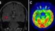
Results: Definitely single lesion abnormality was observed in 35.0% (43/123) of the patients on the MRI portion of PET/MRI. The proportion increased to 74.0% (91/123) when 18 F-FDG PET portion was added. About 75% (69/91) of patients displaying a clear-cut lesion on 18 F-FDG PET/MRI were classified as Engel Class I one-year post-surgery. The proportion of Engel Class I patients was not significantly different when comparing MRI-single lesion patients with MRI-negative, PET-single lesion patients one year after surgery (81.4% vs. 70.0%, P = 0.24). Binary logistic regression analysis revealed that the detection of a clear single lesion on 18 F-FDG PET/MRI was a strong positive predictor of a favorable surgical outcome (OR 3.518, 95% CI 1.363-9.077, p = 0.009).
Conclusion: Single lesion detected on 18 F-FDG PET/MRI is useful to predict good surgical outcome for refractory epilepsy patients; Those patients should be considered as candidates for surgery.
期刊介绍:
Neuroradiology aims to provide state-of-the-art medical and scientific information in the fields of Neuroradiology, Neurosciences, Neurology, Psychiatry, Neurosurgery, and related medical specialities. Neuroradiology as the official Journal of the European Society of Neuroradiology receives submissions from all parts of the world and publishes peer-reviewed original research, comprehensive reviews, educational papers, opinion papers, and short reports on exceptional clinical observations and new technical developments in the field of Neuroimaging and Neurointervention. The journal has subsections for Diagnostic and Interventional Neuroradiology, Advanced Neuroimaging, Paediatric Neuroradiology, Head-Neck-ENT Radiology, Spine Neuroradiology, and for submissions from Japan. Neuroradiology aims to provide new knowledge about and insights into the function and pathology of the human nervous system that may help to better diagnose and treat nervous system diseases. Neuroradiology is a member of the Committee on Publication Ethics (COPE) and follows the COPE core practices. Neuroradiology prefers articles that are free of bias, self-critical regarding limitations, transparent and clear in describing study participants, methods, and statistics, and short in presenting results. Before peer-review all submissions are automatically checked by iThenticate to assess for potential overlap in prior publication.


 分享
分享
 求助内容:
求助内容: 应助结果提醒方式:
 扫码关注我们
