Deep learning-based segmentation in MRI-(immuno)histological examination of myelin and axonal damage in normal-appearing white matter and white matter hyperintensities
Gemma Solé-Guardia, Matthijs Luijten, Esther Janssen, Ruben Visch, Bram Geenen, Benno Küsters, Jurgen A. H. R. Claassen, Geert Litjens, Frank-Erik de Leeuw, Maximilian Wiesmann, Amanda J. Kiliaan
{"title":"Deep learning-based segmentation in MRI-(immuno)histological examination of myelin and axonal damage in normal-appearing white matter and white matter hyperintensities","authors":"Gemma Solé-Guardia, Matthijs Luijten, Esther Janssen, Ruben Visch, Bram Geenen, Benno Küsters, Jurgen A. H. R. Claassen, Geert Litjens, Frank-Erik de Leeuw, Maximilian Wiesmann, Amanda J. Kiliaan","doi":"10.1111/bpa.13301","DOIUrl":null,"url":null,"abstract":"<p>The major vascular cause of dementia is cerebral small vessel disease (SVD). Its diagnosis relies on imaging hallmarks, such as white matter hyperintensities (WMH). WMH present a heterogenous pathology, including myelin and axonal loss. Yet, these might be only the “tip of the iceberg.” Imaging modalities imply that microstructural alterations underlie still normal-appearing white matter (NAWM), preceding the conversion to WMH. Unfortunately, direct pathological characterization of these microstructural alterations affecting myelinated axonal fibers in WMH, and especially NAWM, is still missing. Given that there are no treatments to significantly reduce WMH progression, it is important to extend our knowledge on pathological processes that might already be occurring within NAWM. Staining of myelin with Luxol Fast Blue, while valuable, fails to assess subtle alterations in white matter microstructure. Therefore, we aimed to quantify myelin surrounding axonal fibers and axonal- and microstructural damage in detail by combining (immuno)histochemistry with polarized light imaging (PLI). To study the extent (of early) microstructural damage from periventricular NAWM to the center of WMH, we refined current analysis techniques by using deep learning to define smaller segments of white matter, capturing increasing fluid-attenuated inversion recovery signal. Integration of (immuno)histochemistry and PLI with post-mortem imaging of the brains of individuals with hypertension and normotensive controls enables voxel-wise assessment of the pathology throughout periventricular WMH and NAWM. Myelin loss, axonal integrity, and white matter microstructural damage are not limited to WMH but already occur within NAWM. Notably, we found that axonal damage is higher in individuals with hypertension, particularly in NAWM. These findings highlight the added value of advanced segmentation techniques to visualize subtle changes occurring already in NAWM preceding WMH. By using quantitative MRI and advanced diffusion MRI, future studies may elucidate these very early mechanisms leading to neurodegeneration, which ultimately contribute to the conversion of NAWM to WMH.</p>","PeriodicalId":9290,"journal":{"name":"Brain Pathology","volume":"35 2","pages":""},"PeriodicalIF":6.2000,"publicationDate":"2024-08-23","publicationTypes":"Journal Article","fieldsOfStudy":null,"isOpenAccess":false,"openAccessPdf":"https://onlinelibrary.wiley.com/doi/epdf/10.1111/bpa.13301","citationCount":"0","resultStr":null,"platform":"Semanticscholar","paperid":null,"PeriodicalName":"Brain Pathology","FirstCategoryId":"3","ListUrlMain":"https://onlinelibrary.wiley.com/doi/10.1111/bpa.13301","RegionNum":2,"RegionCategory":"医学","ArticlePicture":[],"TitleCN":null,"AbstractTextCN":null,"PMCID":null,"EPubDate":"","PubModel":"","JCR":"Q1","JCRName":"CLINICAL NEUROLOGY","Score":null,"Total":0}
引用次数: 0
Abstract
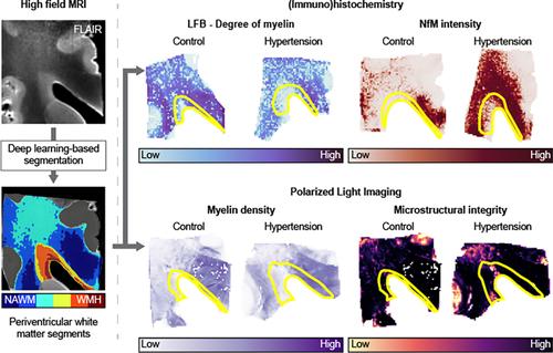
The major vascular cause of dementia is cerebral small vessel disease (SVD). Its diagnosis relies on imaging hallmarks, such as white matter hyperintensities (WMH). WMH present a heterogenous pathology, including myelin and axonal loss. Yet, these might be only the “tip of the iceberg.” Imaging modalities imply that microstructural alterations underlie still normal-appearing white matter (NAWM), preceding the conversion to WMH. Unfortunately, direct pathological characterization of these microstructural alterations affecting myelinated axonal fibers in WMH, and especially NAWM, is still missing. Given that there are no treatments to significantly reduce WMH progression, it is important to extend our knowledge on pathological processes that might already be occurring within NAWM. Staining of myelin with Luxol Fast Blue, while valuable, fails to assess subtle alterations in white matter microstructure. Therefore, we aimed to quantify myelin surrounding axonal fibers and axonal- and microstructural damage in detail by combining (immuno)histochemistry with polarized light imaging (PLI). To study the extent (of early) microstructural damage from periventricular NAWM to the center of WMH, we refined current analysis techniques by using deep learning to define smaller segments of white matter, capturing increasing fluid-attenuated inversion recovery signal. Integration of (immuno)histochemistry and PLI with post-mortem imaging of the brains of individuals with hypertension and normotensive controls enables voxel-wise assessment of the pathology throughout periventricular WMH and NAWM. Myelin loss, axonal integrity, and white matter microstructural damage are not limited to WMH but already occur within NAWM. Notably, we found that axonal damage is higher in individuals with hypertension, particularly in NAWM. These findings highlight the added value of advanced segmentation techniques to visualize subtle changes occurring already in NAWM preceding WMH. By using quantitative MRI and advanced diffusion MRI, future studies may elucidate these very early mechanisms leading to neurodegeneration, which ultimately contribute to the conversion of NAWM to WMH.
期刊介绍:
Brain Pathology is the journal of choice for biomedical scientists investigating diseases of the nervous system. The official journal of the International Society of Neuropathology, Brain Pathology is a peer-reviewed quarterly publication that includes original research, review articles and symposia focuses on the pathogenesis of neurological disease.


 分享
分享
 求助内容:
求助内容: 应助结果提醒方式:
 扫码关注我们
