Oscillatory and nonoscillatory sleep electroencephalographic biomarkers of the epileptic network

IF 6.6 1区 医学 Q1 CLINICAL NEUROLOGY Epilepsia Pub Date : 2024-08-24 DOI:10.1111/epi.18088
Véronique Latreille, Justin Corbin-Lapointe, Laure Peter-Derex, John Thomas, Jean-Marc Lina, Birgit Frauscher
{"title":"Oscillatory and nonoscillatory sleep electroencephalographic biomarkers of the epileptic network","authors":"Véronique Latreille,&nbsp;Justin Corbin-Lapointe,&nbsp;Laure Peter-Derex,&nbsp;John Thomas,&nbsp;Jean-Marc Lina,&nbsp;Birgit Frauscher","doi":"10.1111/epi.18088","DOIUrl":null,"url":null,"abstract":"<div>\n \n \n <section>\n \n <h3> Objective</h3>\n \n <p>In addition to the oscillatory brain activity, the nonoscillatory (scale-free) components of the background electroencephalogram (EEG) may provide further information about the complexity of the underlying neuronal network. As epilepsy is considered a network disease, such scale-free metrics might help to delineate the epileptic network. Here, we performed an analysis of the sleep oscillatory (spindle, slow wave, and rhythmic spectral power) and nonoscillatory (<i>H</i> exponent) intracranial EEG using multiple interictal features to estimate whether and how they deviate from normalcy in 38 adults with drug-resistant epilepsy.</p>\n </section>\n \n <section>\n \n <h3> Methods</h3>\n \n <p>To quantify intracranial EEG abnormalities within and outside the seizure onset areas, patients' values were adjusted based on normative maps derived from the open-access Montreal Neurological Institute open iEEG Atlas. In a subset of 29 patients who underwent resective surgery, we estimated the predictive value of these features to identify the epileptogenic zone in those with a good postsurgical outcome.</p>\n </section>\n \n <section>\n \n <h3> Results</h3>\n \n <p>We found that distinct sleep oscillatory and nonoscillatory metrics behave differently across the epileptic network, with the strongest differences observed for (1) a reduction in spindle activity (spindle rates and rhythmic sigma power in the 10–16 Hz band), (2) a higher rhythmic gamma power (30–80 Hz), and (3) a higher <i>H</i> exponent (steeper 1/<i>f</i> slope). As expected, epileptic spikes were also highest in the seizure onset areas. Furthermore, in surgical patients, the <i>H</i> exponent achieved the highest performance (balanced accuracy of .76) for classifying resected versus nonresected channels in good outcome patients.</p>\n </section>\n \n <section>\n \n <h3> Significance</h3>\n \n <p>This work suggests that nonoscillatory components of the intracranial EEG signal could serve as promising interictal sleep candidates of epileptogenicity in patients with drug-resistant epilepsy. Our findings further advance the understanding of epilepsy as a disease, whereby absence or loss of sleep physiology may provide information complementary to pathological epileptic processes.</p>\n </section>\n </div>","PeriodicalId":11768,"journal":{"name":"Epilepsia","volume":"65 10","pages":"3038-3051"},"PeriodicalIF":6.6000,"publicationDate":"2024-08-24","publicationTypes":"Journal Article","fieldsOfStudy":null,"isOpenAccess":false,"openAccessPdf":"https://onlinelibrary.wiley.com/doi/epdf/10.1111/epi.18088","citationCount":"0","resultStr":null,"platform":"Semanticscholar","paperid":null,"PeriodicalName":"Epilepsia","FirstCategoryId":"3","ListUrlMain":"https://onlinelibrary.wiley.com/doi/10.1111/epi.18088","RegionNum":1,"RegionCategory":"医学","ArticlePicture":[],"TitleCN":null,"AbstractTextCN":null,"PMCID":null,"EPubDate":"","PubModel":"","JCR":"Q1","JCRName":"CLINICAL NEUROLOGY","Score":null,"Total":0}
引用次数: 0

Abstract

Objective

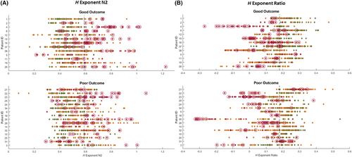
In addition to the oscillatory brain activity, the nonoscillatory (scale-free) components of the background electroencephalogram (EEG) may provide further information about the complexity of the underlying neuronal network. As epilepsy is considered a network disease, such scale-free metrics might help to delineate the epileptic network. Here, we performed an analysis of the sleep oscillatory (spindle, slow wave, and rhythmic spectral power) and nonoscillatory (H exponent) intracranial EEG using multiple interictal features to estimate whether and how they deviate from normalcy in 38 adults with drug-resistant epilepsy.

Methods

To quantify intracranial EEG abnormalities within and outside the seizure onset areas, patients' values were adjusted based on normative maps derived from the open-access Montreal Neurological Institute open iEEG Atlas. In a subset of 29 patients who underwent resective surgery, we estimated the predictive value of these features to identify the epileptogenic zone in those with a good postsurgical outcome.

Results

We found that distinct sleep oscillatory and nonoscillatory metrics behave differently across the epileptic network, with the strongest differences observed for (1) a reduction in spindle activity (spindle rates and rhythmic sigma power in the 10–16 Hz band), (2) a higher rhythmic gamma power (30–80 Hz), and (3) a higher H exponent (steeper 1/f slope). As expected, epileptic spikes were also highest in the seizure onset areas. Furthermore, in surgical patients, the H exponent achieved the highest performance (balanced accuracy of .76) for classifying resected versus nonresected channels in good outcome patients.

Significance

This work suggests that nonoscillatory components of the intracranial EEG signal could serve as promising interictal sleep candidates of epileptogenicity in patients with drug-resistant epilepsy. Our findings further advance the understanding of epilepsy as a disease, whereby absence or loss of sleep physiology may provide information complementary to pathological epileptic processes.
Abstract Image

查看原文
分享 分享
微信好友 朋友圈 QQ好友 复制链接
本刊更多论文
癫痫网络的振荡性和非振荡性睡眠脑电图生物标志物。
目的:除大脑振荡活动外,背景脑电图(EEG)中的非振荡(无标度)成分也可提供有关潜在神经元网络复杂性的进一步信息。由于癫痫被认为是一种网络疾病,这种无标度指标可能有助于划分癫痫网络。在此,我们利用多种发作间期特征对睡眠振荡(纺锤波、慢波和节律频谱功率)和非振荡(H 指数)颅内脑电图进行了分析,以估计 38 名成人耐药性癫痫患者的睡眠振荡和非振荡脑电图是否偏离正常以及如何偏离正常:为了量化发作起始区内外的颅内脑电图异常,根据开放访问的蒙特利尔神经研究所开放 iEEG 图谱得出的常模图调整了患者的数值。在接受切除手术的 29 例患者中,我们估算了这些特征的预测价值,以确定手术后疗效良好的患者的致痫区:我们发现,不同的睡眠振荡和非振荡指标在整个癫痫网络中的表现各不相同,其中差异最大的是:(1) 纺锤体活动的减少(10-16 Hz 频段的纺锤体率和节律性 sigma 功率);(2) 更高的节律性 gamma 功率(30-80 Hz);(3) 更高的 H 指数(更陡的 1/f 斜坡)。不出所料,癫痫发作区的癫痫尖峰也是最高的。此外,在手术患者中,H 指数在对结果良好患者的切除与未切除通道进行分类时达到了最高的性能(平衡准确率为 0.76):这项研究表明,颅内脑电信号的非振荡成分可作为耐药性癫痫患者发作间期睡眠致痫性的候选成分。我们的研究结果进一步加深了人们对癫痫这种疾病的认识,睡眠生理缺失或丧失可提供病理癫痫过程的补充信息。
本文章由计算机程序翻译,如有差异,请以英文原文为准。
求助全文
约1分钟内获得全文 去求助
来源期刊
Epilepsia
Epilepsia 医学-临床神经学
CiteScore
10.90
自引率
10.70%
发文量
319
审稿时长
2-4 weeks
期刊介绍: Epilepsia is the leading, authoritative source for innovative clinical and basic science research for all aspects of epilepsy and seizures. In addition, Epilepsia publishes critical reviews, opinion pieces, and guidelines that foster understanding and aim to improve the diagnosis and treatment of people with seizures and epilepsy.
期刊最新文献
Structural connectivity changes in focal epilepsy: Beyond the epileptogenic zone. Efficacy and safety of perampanel in patients with seizures associated with Lennox-Gastaut syndrome: A randomized trial. Focal negative motor seizures: Multimodal evaluation. Automatic responsiveness testing in epilepsy with wearable technology: The ARTiE Watch. WONOEP appraisal: Targeted therapy development for early onset epilepsies.
×
引用
GB/T 7714-2015
复制
MLA
复制
APA
复制
导出至
BibTeX EndNote RefMan NoteFirst NoteExpress
×
×
提示
您的信息不完整,为了账户安全,请先补充。
现在去补充
×
提示
您因"违规操作"
具体请查看互助需知
我知道了
×
提示
现在去查看 取消
×
提示
确定
0
微信
客服QQ
Book学术公众号 扫码关注我们
反馈
×
意见反馈
请填写您的意见或建议
请填写您的手机或邮箱
已复制链接
已复制链接
快去分享给好友吧!
我知道了
×
扫码分享
扫码分享
Book学术官方微信
Book学术文献互助
Book学术文献互助群
群 号:481959085
Book学术
文献互助 智能选刊 最新文献 互助须知 联系我们:info@booksci.cn
Book学术提供免费学术资源搜索服务,方便国内外学者检索中英文文献。致力于提供最便捷和优质的服务体验。
Copyright © 2023 Book学术 All rights reserved.
ghs 京公网安备 11010802042870号 京ICP备2023020795号-1