Paweł Łajczak, Jakub Matyja, Kamil Jóźwik, Zbigniew Nawrat
{"title":"Accuracy of vestibular schwannoma segmentation using deep learning models - a systematic review & meta-analysis.","authors":"Paweł Łajczak, Jakub Matyja, Kamil Jóźwik, Zbigniew Nawrat","doi":"10.1007/s00234-024-03449-1","DOIUrl":null,"url":null,"abstract":"<p><p>Vestibular Schwannoma (VS) is a rare tumor with varied incidence rates, predominantly affecting the 60-69 age group. In the era of artificial intelligence (AI), deep learning (DL) algorithms show promise in automating diagnosis. However, a knowledge gap exists in the automated segmentation of VS using DL. To address this gap, this meta-analysis aims to provide insights into the current state of DL algorithms applied to MR images of VS.</p><p><strong>Methodology: </strong>Following 2020 PRISMA guidelines, a search across four databases was conducted. Inclusion criteria focused on articles using DL for VS MR image segmentation. The primary metric was the Dice score, supplemented by relative volume error (RVE) and average symmetric surface distance (ASSD).</p><p><strong>Results: </strong>The search process identified 752 articles, leading to 11 studies for meta-analysis. A QUADAS- 2 analysis revealed varying biases. The overall Dice score for 56 models was 0.89 (CI: 0.88-0.90), with high heterogeneity (I2 = 95.9%). Subgroup analyses based on DL architecture, MRI inputs, and testing set sizes revealed performance variations. 2.5D DL networks demonstrated comparable efficacy to 3D networks. Imaging input analyses highlighted the superiority of contrast-enhanced T1-weighted imaging and mixed MRI inputs.</p><p><strong>Discussion: </strong>This study fills a gap in systematic review in the automated segmentation of VS using DL techniques. Despite promising results, limitations include publication bias and high heterogeneity. Future research should focus on standardized designs, larger testing sets, and addressing biases for more reliable results. DL have promising efficacy in VS diagnosis, however further validation and standardization is needed.</p><p><strong>Conclusion: </strong>In conclusion, this meta-analysis provides comprehensive review into the current landscape of automated VS segmentation using DL. The high Dice score indicates promising agreement in segmentation, yet challenges like bias and heterogeneity must be addressed in the future research.</p>","PeriodicalId":19422,"journal":{"name":"Neuroradiology","volume":" ","pages":"729-742"},"PeriodicalIF":2.6000,"publicationDate":"2025-03-01","publicationTypes":"Journal Article","fieldsOfStudy":null,"isOpenAccess":false,"openAccessPdf":"https://www.ncbi.nlm.nih.gov/pmc/articles/PMC12003617/pdf/","citationCount":"0","resultStr":null,"platform":"Semanticscholar","paperid":null,"PeriodicalName":"Neuroradiology","FirstCategoryId":"3","ListUrlMain":"https://doi.org/10.1007/s00234-024-03449-1","RegionNum":3,"RegionCategory":"医学","ArticlePicture":[],"TitleCN":null,"AbstractTextCN":null,"PMCID":null,"EPubDate":"2024/8/24 0:00:00","PubModel":"Epub","JCR":"Q2","JCRName":"CLINICAL NEUROLOGY","Score":null,"Total":0}
引用次数: 0
Abstract
Vestibular Schwannoma (VS) is a rare tumor with varied incidence rates, predominantly affecting the 60-69 age group. In the era of artificial intelligence (AI), deep learning (DL) algorithms show promise in automating diagnosis. However, a knowledge gap exists in the automated segmentation of VS using DL. To address this gap, this meta-analysis aims to provide insights into the current state of DL algorithms applied to MR images of VS.
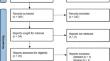
Methodology: Following 2020 PRISMA guidelines, a search across four databases was conducted. Inclusion criteria focused on articles using DL for VS MR image segmentation. The primary metric was the Dice score, supplemented by relative volume error (RVE) and average symmetric surface distance (ASSD).
Results: The search process identified 752 articles, leading to 11 studies for meta-analysis. A QUADAS- 2 analysis revealed varying biases. The overall Dice score for 56 models was 0.89 (CI: 0.88-0.90), with high heterogeneity (I2 = 95.9%). Subgroup analyses based on DL architecture, MRI inputs, and testing set sizes revealed performance variations. 2.5D DL networks demonstrated comparable efficacy to 3D networks. Imaging input analyses highlighted the superiority of contrast-enhanced T1-weighted imaging and mixed MRI inputs.
Discussion: This study fills a gap in systematic review in the automated segmentation of VS using DL techniques. Despite promising results, limitations include publication bias and high heterogeneity. Future research should focus on standardized designs, larger testing sets, and addressing biases for more reliable results. DL have promising efficacy in VS diagnosis, however further validation and standardization is needed.
Conclusion: In conclusion, this meta-analysis provides comprehensive review into the current landscape of automated VS segmentation using DL. The high Dice score indicates promising agreement in segmentation, yet challenges like bias and heterogeneity must be addressed in the future research.
期刊介绍:
Neuroradiology aims to provide state-of-the-art medical and scientific information in the fields of Neuroradiology, Neurosciences, Neurology, Psychiatry, Neurosurgery, and related medical specialities. Neuroradiology as the official Journal of the European Society of Neuroradiology receives submissions from all parts of the world and publishes peer-reviewed original research, comprehensive reviews, educational papers, opinion papers, and short reports on exceptional clinical observations and new technical developments in the field of Neuroimaging and Neurointervention. The journal has subsections for Diagnostic and Interventional Neuroradiology, Advanced Neuroimaging, Paediatric Neuroradiology, Head-Neck-ENT Radiology, Spine Neuroradiology, and for submissions from Japan. Neuroradiology aims to provide new knowledge about and insights into the function and pathology of the human nervous system that may help to better diagnose and treat nervous system diseases. Neuroradiology is a member of the Committee on Publication Ethics (COPE) and follows the COPE core practices. Neuroradiology prefers articles that are free of bias, self-critical regarding limitations, transparent and clear in describing study participants, methods, and statistics, and short in presenting results. Before peer-review all submissions are automatically checked by iThenticate to assess for potential overlap in prior publication.


 分享
分享
 求助内容:
求助内容: 应助结果提醒方式:
 扫码关注我们
