Ortrud Uckermann, Jonathan Ziegler, Matthias Meinhardt, Sven Richter, Gabriele Schackert, Ilker Y Eyüpoglu, Mido M Hijazi, Dietmar Krex, Tareq A Juratli, Stephan B Sobottka, Roberta Galli
{"title":"Raman and autofluorescence spectroscopy for in situ identification of neoplastic tissue during surgical treatment of brain tumors.","authors":"Ortrud Uckermann, Jonathan Ziegler, Matthias Meinhardt, Sven Richter, Gabriele Schackert, Ilker Y Eyüpoglu, Mido M Hijazi, Dietmar Krex, Tareq A Juratli, Stephan B Sobottka, Roberta Galli","doi":"10.1007/s11060-024-04809-w","DOIUrl":null,"url":null,"abstract":"<p><strong>Purpose: </strong>Raman spectroscopy (RS) is a promising method for brain tumor detection. Near-infrared autofluorescence (AF) acquired during RS provides additional useful information for tumor identification and was investigated in comparison with RS for delineating brain tumors in situ.</p><p><strong>Methods: </strong>Raman spectra were acquired together with AF in situ within the solid tumor and at the tumor border during routine brain tumor surgeries (218 spectra; glioma WHO II-III, n = 6; GBM, n = 10; metastases, n = 10; meningioma, n = 3). Tissue classification for tumor identification in situ was trained on ex vivo data (375 spectra; glioma/GBM patients, n = 20; metastases, n = 11; meningioma, n = 13; and epileptic hippocampi, n = 4).</p><p><strong>Results: </strong>Both in situ and ex vivo data showed that AF intensity in brain tumors was lower than that in border regions and normal brain tissue. Moreover, a positive correlation was observed between the AF intensity and the intensity of the Raman band corresponding to lipids at 1437 cm<sup>- 1</sup>, while a negative correlation was found with the intensity of the protein band at 1260 cm<sup>- 1</sup>. The classification of in situ AF and RS datasets matched the surgeon's evaluation of tissue type, with correct rates of 0.83 and 0.84, respectively. Similar correct rates were achieved in comparison to histopathology of tissue biopsies resected in selected measurement positions (AF: 0.80, RS: 0.83).</p><p><strong>Conclusions: </strong>Spectroscopy was successfully integrated into existing neurosurgical workflows, and in situ spectroscopic data could be classified based on ex vivo data. RS confirmed its ability to detect brain tumors, while AF emerged as a competitive method for intraoperative tumor delineation.</p>","PeriodicalId":16425,"journal":{"name":"Journal of Neuro-Oncology","volume":" ","pages":"543-553"},"PeriodicalIF":3.1000,"publicationDate":"2024-12-01","publicationTypes":"Journal Article","fieldsOfStudy":null,"isOpenAccess":false,"openAccessPdf":"https://www.ncbi.nlm.nih.gov/pmc/articles/PMC11614956/pdf/","citationCount":"0","resultStr":null,"platform":"Semanticscholar","paperid":null,"PeriodicalName":"Journal of Neuro-Oncology","FirstCategoryId":"3","ListUrlMain":"https://doi.org/10.1007/s11060-024-04809-w","RegionNum":2,"RegionCategory":"医学","ArticlePicture":[],"TitleCN":null,"AbstractTextCN":null,"PMCID":null,"EPubDate":"2024/8/28 0:00:00","PubModel":"Epub","JCR":"Q2","JCRName":"CLINICAL NEUROLOGY","Score":null,"Total":0}
引用次数: 0
Abstract
Purpose: Raman spectroscopy (RS) is a promising method for brain tumor detection. Near-infrared autofluorescence (AF) acquired during RS provides additional useful information for tumor identification and was investigated in comparison with RS for delineating brain tumors in situ.
Methods: Raman spectra were acquired together with AF in situ within the solid tumor and at the tumor border during routine brain tumor surgeries (218 spectra; glioma WHO II-III, n = 6; GBM, n = 10; metastases, n = 10; meningioma, n = 3). Tissue classification for tumor identification in situ was trained on ex vivo data (375 spectra; glioma/GBM patients, n = 20; metastases, n = 11; meningioma, n = 13; and epileptic hippocampi, n = 4).
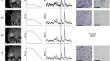
Results: Both in situ and ex vivo data showed that AF intensity in brain tumors was lower than that in border regions and normal brain tissue. Moreover, a positive correlation was observed between the AF intensity and the intensity of the Raman band corresponding to lipids at 1437 cm- 1, while a negative correlation was found with the intensity of the protein band at 1260 cm- 1. The classification of in situ AF and RS datasets matched the surgeon's evaluation of tissue type, with correct rates of 0.83 and 0.84, respectively. Similar correct rates were achieved in comparison to histopathology of tissue biopsies resected in selected measurement positions (AF: 0.80, RS: 0.83).
Conclusions: Spectroscopy was successfully integrated into existing neurosurgical workflows, and in situ spectroscopic data could be classified based on ex vivo data. RS confirmed its ability to detect brain tumors, while AF emerged as a competitive method for intraoperative tumor delineation.
期刊介绍:
The Journal of Neuro-Oncology is a multi-disciplinary journal encompassing basic, applied, and clinical investigations in all research areas as they relate to cancer and the central nervous system. It provides a single forum for communication among neurologists, neurosurgeons, radiotherapists, medical oncologists, neuropathologists, neurodiagnosticians, and laboratory-based oncologists conducting relevant research. The Journal of Neuro-Oncology does not seek to isolate the field, but rather to focus the efforts of many disciplines in one publication through a format which pulls together these diverse interests. More than any other field of oncology, cancer of the central nervous system requires multi-disciplinary approaches. To alleviate having to scan dozens of journals of cell biology, pathology, laboratory and clinical endeavours, JNO is a periodical in which current, high-quality, relevant research in all aspects of neuro-oncology may be found.


 分享
分享
 求助内容:
求助内容: 应助结果提醒方式:
 扫码关注我们
