{"title":"Deep Learning-Assisted Automatic Diagnosis of Anterior Cruciate Ligament Tear in Knee Magnetic Resonance Images.","authors":"Xuanwei Wang, Yuanfeng Wu, Jiafeng Li, Yifan Li, Sanzhong Xu","doi":"10.3390/tomography10080094","DOIUrl":null,"url":null,"abstract":"<p><p>Anterior cruciate ligament (ACL) tears are prevalent knee injures, particularly among active individuals. Accurate and timely diagnosis is essential for determining the optimal treatment strategy and assessing patient prognosis. Various previous studies have demonstrated the successful application of deep learning techniques in the field of medical image analysis. This study aimed to develop a deep learning model for detecting ACL tears in knee magnetic resonance Imaging (MRI) to enhance diagnostic accuracy and efficiency. The proposed model consists of three main modules: a Dual-Scale Data Augmentation module (DDA) to enrich the training data on both the spatial and layer scales; a selective group attention module (SG) to capture relationships across the layer, channel, and space scales; and a fusion module to explore the inter-relationships among various perspectives to achieve the final classification. To ensure a fair comparison, the study utilized a public dataset from MRNet, comprising knee MRI scans from 1250 exams, with a focus on three distinct views: axial, coronal, and sagittal. The experimental results demonstrate the superior performance of the proposed model, termed SGNET, in ACL tear detection compared with other comparison models, achieving an accuracy of 0.9250, a sensitivity of 0.9259, a specificity of 0.9242, and an AUC of 0.9747.</p>","PeriodicalId":51330,"journal":{"name":"Tomography","volume":"10 8","pages":"1263-1276"},"PeriodicalIF":2.2000,"publicationDate":"2024-08-13","publicationTypes":"Journal Article","fieldsOfStudy":null,"isOpenAccess":false,"openAccessPdf":"https://www.ncbi.nlm.nih.gov/pmc/articles/PMC11487377/pdf/","citationCount":"0","resultStr":null,"platform":"Semanticscholar","paperid":null,"PeriodicalName":"Tomography","FirstCategoryId":"3","ListUrlMain":"https://doi.org/10.3390/tomography10080094","RegionNum":4,"RegionCategory":"医学","ArticlePicture":[],"TitleCN":null,"AbstractTextCN":null,"PMCID":null,"EPubDate":"","PubModel":"","JCR":"Q2","JCRName":"RADIOLOGY, NUCLEAR MEDICINE & MEDICAL IMAGING","Score":null,"Total":0}
引用次数: 0
Abstract
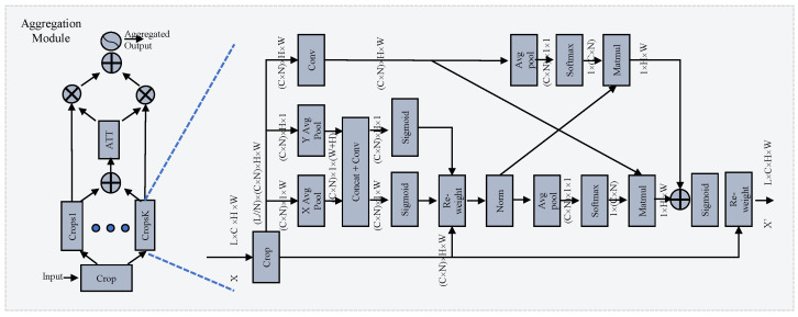
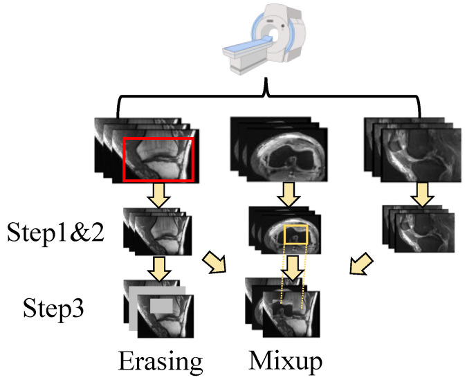
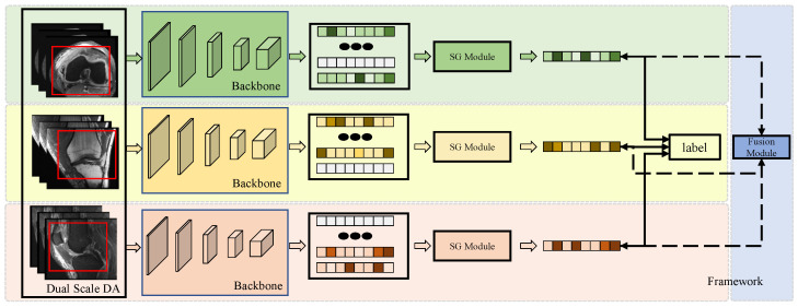
Anterior cruciate ligament (ACL) tears are prevalent knee injures, particularly among active individuals. Accurate and timely diagnosis is essential for determining the optimal treatment strategy and assessing patient prognosis. Various previous studies have demonstrated the successful application of deep learning techniques in the field of medical image analysis. This study aimed to develop a deep learning model for detecting ACL tears in knee magnetic resonance Imaging (MRI) to enhance diagnostic accuracy and efficiency. The proposed model consists of three main modules: a Dual-Scale Data Augmentation module (DDA) to enrich the training data on both the spatial and layer scales; a selective group attention module (SG) to capture relationships across the layer, channel, and space scales; and a fusion module to explore the inter-relationships among various perspectives to achieve the final classification. To ensure a fair comparison, the study utilized a public dataset from MRNet, comprising knee MRI scans from 1250 exams, with a focus on three distinct views: axial, coronal, and sagittal. The experimental results demonstrate the superior performance of the proposed model, termed SGNET, in ACL tear detection compared with other comparison models, achieving an accuracy of 0.9250, a sensitivity of 0.9259, a specificity of 0.9242, and an AUC of 0.9747.
TomographyMedicine-Radiology, Nuclear Medicine and Imaging
CiteScore
2.70
自引率
10.50%
发文量
222
期刊介绍:
TomographyTM publishes basic (technical and pre-clinical) and clinical scientific articles which involve the advancement of imaging technologies. Tomography encompasses studies that use single or multiple imaging modalities including for example CT, US, PET, SPECT, MR and hyperpolarization technologies, as well as optical modalities (i.e. bioluminescence, photoacoustic, endomicroscopy, fiber optic imaging and optical computed tomography) in basic sciences, engineering, preclinical and clinical medicine.
Tomography also welcomes studies involving exploration and refinement of contrast mechanisms and image-derived metrics within and across modalities toward the development of novel imaging probes for image-based feedback and intervention. The use of imaging in biology and medicine provides unparalleled opportunities to noninvasively interrogate tissues to obtain real-time dynamic and quantitative information required for diagnosis and response to interventions and to follow evolving pathological conditions. As multi-modal studies and the complexities of imaging technologies themselves are ever increasing to provide advanced information to scientists and clinicians.
Tomography provides a unique publication venue allowing investigators the opportunity to more precisely communicate integrated findings related to the diverse and heterogeneous features associated with underlying anatomical, physiological, functional, metabolic and molecular genetic activities of normal and diseased tissue. Thus Tomography publishes peer-reviewed articles which involve the broad use of imaging of any tissue and disease type including both preclinical and clinical investigations. In addition, hardware/software along with chemical and molecular probe advances are welcome as they are deemed to significantly contribute towards the long-term goal of improving the overall impact of imaging on scientific and clinical discovery.


 分享
分享
 求助内容:
求助内容: 应助结果提醒方式:
 扫码关注我们
