Shingo Kato, Mai Azuma, Nobuyuki Horita, Daisuke Utsunomiya
{"title":"Monitoring the Efficacy of Tafamidis in ATTR Cardiac Amyloidosis by MRI-ECV: A Systematic Review and Meta-Analysis.","authors":"Shingo Kato, Mai Azuma, Nobuyuki Horita, Daisuke Utsunomiya","doi":"10.3390/tomography10080097","DOIUrl":null,"url":null,"abstract":"<p><strong>Background: </strong>The usefulness of monitoring treatment effect of tafamidis using magnetic resonance imaging (MRI) extracellular volume fraction (ECV) has been reported.</p><p><strong>Objective: </strong>we conducted a meta-analysis to evaluate the usefulness of this method.</p><p><strong>Methods: </strong>Data from 246 ATTR-CMs from six studies were extracted and included in the analysis. An inverse variance meta-analysis using a random effects model was performed to evaluate the change in MRI-ECV before and after tafamidis treatment. The analysis was also performed by classifying the patients into ATTR-CM types (wild-type or hereditary).</p><p><strong>Results: </strong>ECV change before and after tafamidis treatment was 0.33% (95% CI: -1.83-2.49, I<sup>2</sup> = 0%, p = 0.76 for heterogeneity) in the treatment group and 4.23% (95% CI: 0.44-8.02, I<sup>2</sup> = 0%, p = 0.18 for heterogeneity) in the non-treatment group. The change in ECV before and after treatment was not significant in the treated group (p = 0.76), but there was a significant increase in the non-treated group (p = 0.03). There was no difference in the change in ECV between wild-type (95% CI: -2.65-3.40) and hereditary-type (95% CI: -9.28-4.28) (p = 0.45).</p><p><strong>Conclusions: </strong>The results of this meta-analysis suggest that MRI-ECV measurement is a useful imaging method for noninvasively evaluating the efficacy of tafamidis treatment for ATTR-CM.</p>","PeriodicalId":51330,"journal":{"name":"Tomography","volume":"10 8","pages":"1303-1311"},"PeriodicalIF":2.2000,"publicationDate":"2024-08-16","publicationTypes":"Journal Article","fieldsOfStudy":null,"isOpenAccess":false,"openAccessPdf":"https://www.ncbi.nlm.nih.gov/pmc/articles/PMC11360159/pdf/","citationCount":"0","resultStr":null,"platform":"Semanticscholar","paperid":null,"PeriodicalName":"Tomography","FirstCategoryId":"3","ListUrlMain":"https://doi.org/10.3390/tomography10080097","RegionNum":4,"RegionCategory":"医学","ArticlePicture":[],"TitleCN":null,"AbstractTextCN":null,"PMCID":null,"EPubDate":"","PubModel":"","JCR":"Q2","JCRName":"RADIOLOGY, NUCLEAR MEDICINE & MEDICAL IMAGING","Score":null,"Total":0}
引用次数: 0
Abstract
Background: The usefulness of monitoring treatment effect of tafamidis using magnetic resonance imaging (MRI) extracellular volume fraction (ECV) has been reported.
Objective: we conducted a meta-analysis to evaluate the usefulness of this method.
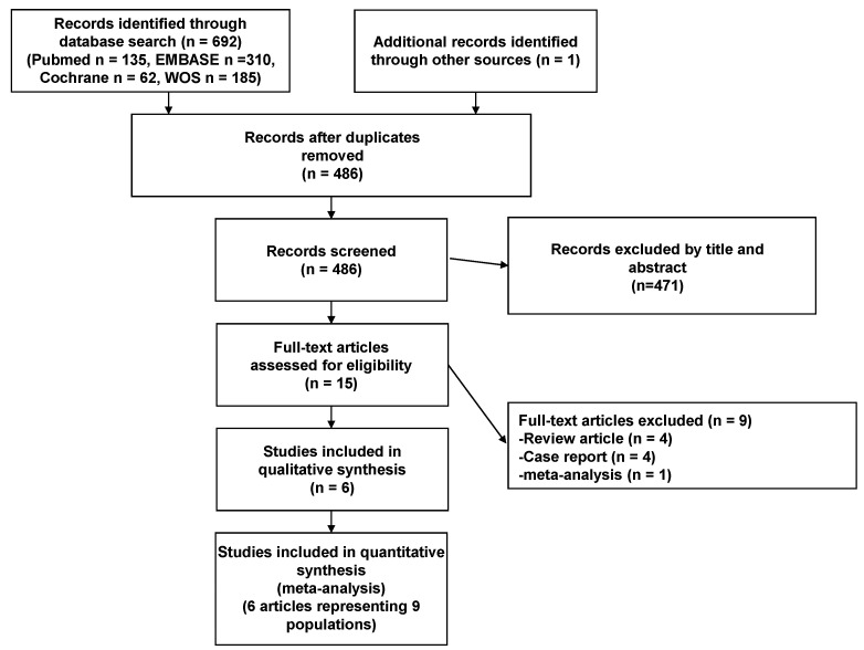
Methods: Data from 246 ATTR-CMs from six studies were extracted and included in the analysis. An inverse variance meta-analysis using a random effects model was performed to evaluate the change in MRI-ECV before and after tafamidis treatment. The analysis was also performed by classifying the patients into ATTR-CM types (wild-type or hereditary).
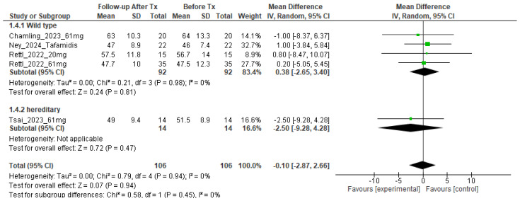
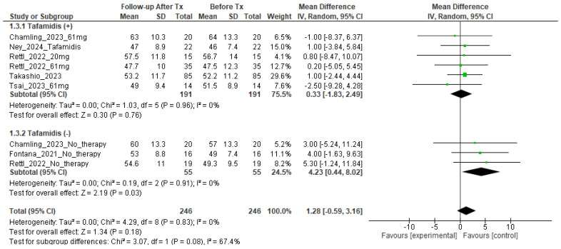
Results: ECV change before and after tafamidis treatment was 0.33% (95% CI: -1.83-2.49, I2 = 0%, p = 0.76 for heterogeneity) in the treatment group and 4.23% (95% CI: 0.44-8.02, I2 = 0%, p = 0.18 for heterogeneity) in the non-treatment group. The change in ECV before and after treatment was not significant in the treated group (p = 0.76), but there was a significant increase in the non-treated group (p = 0.03). There was no difference in the change in ECV between wild-type (95% CI: -2.65-3.40) and hereditary-type (95% CI: -9.28-4.28) (p = 0.45).
Conclusions: The results of this meta-analysis suggest that MRI-ECV measurement is a useful imaging method for noninvasively evaluating the efficacy of tafamidis treatment for ATTR-CM.
TomographyMedicine-Radiology, Nuclear Medicine and Imaging
CiteScore
2.70
自引率
10.50%
发文量
222
期刊介绍:
TomographyTM publishes basic (technical and pre-clinical) and clinical scientific articles which involve the advancement of imaging technologies. Tomography encompasses studies that use single or multiple imaging modalities including for example CT, US, PET, SPECT, MR and hyperpolarization technologies, as well as optical modalities (i.e. bioluminescence, photoacoustic, endomicroscopy, fiber optic imaging and optical computed tomography) in basic sciences, engineering, preclinical and clinical medicine.
Tomography also welcomes studies involving exploration and refinement of contrast mechanisms and image-derived metrics within and across modalities toward the development of novel imaging probes for image-based feedback and intervention. The use of imaging in biology and medicine provides unparalleled opportunities to noninvasively interrogate tissues to obtain real-time dynamic and quantitative information required for diagnosis and response to interventions and to follow evolving pathological conditions. As multi-modal studies and the complexities of imaging technologies themselves are ever increasing to provide advanced information to scientists and clinicians.
Tomography provides a unique publication venue allowing investigators the opportunity to more precisely communicate integrated findings related to the diverse and heterogeneous features associated with underlying anatomical, physiological, functional, metabolic and molecular genetic activities of normal and diseased tissue. Thus Tomography publishes peer-reviewed articles which involve the broad use of imaging of any tissue and disease type including both preclinical and clinical investigations. In addition, hardware/software along with chemical and molecular probe advances are welcome as they are deemed to significantly contribute towards the long-term goal of improving the overall impact of imaging on scientific and clinical discovery.


 分享
分享
 求助内容:
求助内容: 应助结果提醒方式:
 扫码关注我们
