Carmelo Pirri, Nina Pirri, Veronica Macchi, Andrea Porzionato, Raffaele De Caro, Carla Stecco
{"title":"Ultrasound Imaging of Ankle Retinacula: A Comprehensive Review.","authors":"Carmelo Pirri, Nina Pirri, Veronica Macchi, Andrea Porzionato, Raffaele De Caro, Carla Stecco","doi":"10.3390/tomography10080095","DOIUrl":null,"url":null,"abstract":"<p><p>The retinacula of the ankle are specialized anatomical structures characterized by localized thickenings of the crural fascia that envelop the deep components of the lower leg, ankle and foot. The ankle retinacula include the extensor retinacula, the peroneal retinacula and flexor retinaculum. Despite their potential to explain persistent and unexplained pain following an injury, these structures are often overlooked or incorrectly diagnosed. Hence, this comprehensive review was performed aiming to investigate the use and the methodology of US imaging to assess ankle retinacula. The search was performed on PubMed and Web of Science databases from inception to May 2024. The MeSH keywords used were as follows: \"Ankle Retinacula\", \"Foot Retinacula\", \"Superior extensor retinaculum\", \"Inferior extensor retinaculum\", \"peroneal retinaculum\", \"superior peroneal retinaculum\", \"inferior peroneal retinaculum\", \"flexor retinaculum\", \"Ultrasound Imaging\", \"Ultrasound\", \"Ultrasonography\" and \"Ultrasound examination\". In total, 257 records underwent screening, resulting in 22 studies meeting the criteria for inclusion after the process of revision. Data heterogeneity prevents synthesis and consistent conclusions. The results showed that advanced US imaging holds promise as a crucial tool to perform an US examination of ankle retinacula, offering static and dynamic insights into ankle retinacula pathology. Understanding normal anatomy and US imaging is essential for accurately identifying injuries. Future research should focus on clinical trials to validate parameters and ensure their reliability in clinical practice.</p>","PeriodicalId":51330,"journal":{"name":"Tomography","volume":"10 8","pages":"1277-1293"},"PeriodicalIF":2.2000,"publicationDate":"2024-08-14","publicationTypes":"Journal Article","fieldsOfStudy":null,"isOpenAccess":false,"openAccessPdf":"https://www.ncbi.nlm.nih.gov/pmc/articles/PMC11359401/pdf/","citationCount":"0","resultStr":null,"platform":"Semanticscholar","paperid":null,"PeriodicalName":"Tomography","FirstCategoryId":"3","ListUrlMain":"https://doi.org/10.3390/tomography10080095","RegionNum":4,"RegionCategory":"医学","ArticlePicture":[],"TitleCN":null,"AbstractTextCN":null,"PMCID":null,"EPubDate":"","PubModel":"","JCR":"Q2","JCRName":"RADIOLOGY, NUCLEAR MEDICINE & MEDICAL IMAGING","Score":null,"Total":0}
引用次数: 0
Abstract
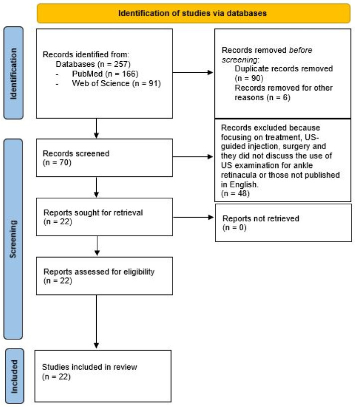
The retinacula of the ankle are specialized anatomical structures characterized by localized thickenings of the crural fascia that envelop the deep components of the lower leg, ankle and foot. The ankle retinacula include the extensor retinacula, the peroneal retinacula and flexor retinaculum. Despite their potential to explain persistent and unexplained pain following an injury, these structures are often overlooked or incorrectly diagnosed. Hence, this comprehensive review was performed aiming to investigate the use and the methodology of US imaging to assess ankle retinacula. The search was performed on PubMed and Web of Science databases from inception to May 2024. The MeSH keywords used were as follows: "Ankle Retinacula", "Foot Retinacula", "Superior extensor retinaculum", "Inferior extensor retinaculum", "peroneal retinaculum", "superior peroneal retinaculum", "inferior peroneal retinaculum", "flexor retinaculum", "Ultrasound Imaging", "Ultrasound", "Ultrasonography" and "Ultrasound examination". In total, 257 records underwent screening, resulting in 22 studies meeting the criteria for inclusion after the process of revision. Data heterogeneity prevents synthesis and consistent conclusions. The results showed that advanced US imaging holds promise as a crucial tool to perform an US examination of ankle retinacula, offering static and dynamic insights into ankle retinacula pathology. Understanding normal anatomy and US imaging is essential for accurately identifying injuries. Future research should focus on clinical trials to validate parameters and ensure their reliability in clinical practice.
踝关节蛛网膜是一种特殊的解剖结构,其特点是嵴状筋膜局部增厚,包裹着小腿、踝关节和足部的深层组织。踝关节蛛网膜包括伸肌蛛网膜、腓肠肌蛛网膜和屈肌蛛网膜。尽管这些结构有可能解释受伤后的持续性不明原因疼痛,但却经常被忽视或误诊。因此,本综述旨在研究使用 US 成像评估踝关节蛛网膜的方法。检索在 PubMed 和 Web of Science 数据库中进行,检索时间从开始到 2024 年 5 月。使用的 MeSH 关键词如下:"踝网膜"、"足网膜"、"上伸肌网膜"、"下伸肌网膜"、"腓网膜"、"上腓网膜"、"下腓网膜"、"屈网膜"、"超声成像"、"超声"、"超声检查 "和 "超声检查"。共有 257 条记录经过筛选,经过修改后有 22 项研究符合纳入标准。数据的异质性妨碍了研究结果的综合和结论的一致性。研究结果表明,先进的 US 成像技术有望成为对踝关节视网膜进行 US 检查的重要工具,为踝关节视网膜病理学提供静态和动态的见解。了解正常解剖结构和 US 成像对准确识别损伤至关重要。未来的研究应侧重于临床试验,以验证参数并确保其在临床实践中的可靠性。
TomographyMedicine-Radiology, Nuclear Medicine and Imaging
CiteScore
2.70
自引率
10.50%
发文量
222
期刊介绍:
TomographyTM publishes basic (technical and pre-clinical) and clinical scientific articles which involve the advancement of imaging technologies. Tomography encompasses studies that use single or multiple imaging modalities including for example CT, US, PET, SPECT, MR and hyperpolarization technologies, as well as optical modalities (i.e. bioluminescence, photoacoustic, endomicroscopy, fiber optic imaging and optical computed tomography) in basic sciences, engineering, preclinical and clinical medicine.
Tomography also welcomes studies involving exploration and refinement of contrast mechanisms and image-derived metrics within and across modalities toward the development of novel imaging probes for image-based feedback and intervention. The use of imaging in biology and medicine provides unparalleled opportunities to noninvasively interrogate tissues to obtain real-time dynamic and quantitative information required for diagnosis and response to interventions and to follow evolving pathological conditions. As multi-modal studies and the complexities of imaging technologies themselves are ever increasing to provide advanced information to scientists and clinicians.
Tomography provides a unique publication venue allowing investigators the opportunity to more precisely communicate integrated findings related to the diverse and heterogeneous features associated with underlying anatomical, physiological, functional, metabolic and molecular genetic activities of normal and diseased tissue. Thus Tomography publishes peer-reviewed articles which involve the broad use of imaging of any tissue and disease type including both preclinical and clinical investigations. In addition, hardware/software along with chemical and molecular probe advances are welcome as they are deemed to significantly contribute towards the long-term goal of improving the overall impact of imaging on scientific and clinical discovery.


 分享
分享
 求助内容:
求助内容: 应助结果提醒方式:
 扫码关注我们
