Katherine E. Schwetye, Lakshmi Ramachandran Nair, Joseph Boyle, Jed A. Barash
{"title":"Histopathologic correlates of opioid-associated injury in CHANTER syndrome: first report of a post-mortem examination","authors":"Katherine E. Schwetye, Lakshmi Ramachandran Nair, Joseph Boyle, Jed A. Barash","doi":"10.1007/s00401-024-02797-9","DOIUrl":null,"url":null,"abstract":"<div><p>Opioid-associated brain injury may involve selective regions, including the hippocampi alone, globi pallidi, and cerebellar hemispheres. Opioid-associated amnestic syndrome, for example, is one clinical correlate of hippocampal injury as manifest by MRI abnormality. When all three regions are involved in what may be a more fulminant injury, the syndrome is termed “cerebellar, hippocampal, and basal nuclei transient edema with restricted diffusion (CHANTER)”, initially described in 2019. Until now, to our knowledge, there have been no histopathologic correlates to the imaging findings specifically in CHANTER syndrome. Here, for the first time, we present histopathologic findings of the post-mortem brain from a patient who died from complications of CHANTER syndrome following fentanyl intoxication. These observations included microhemorrhage, reactive and necrotic vasculature, eosinophilic neuronal necrosis, axonal swelling and spheroids, and frank infarction. The findings support previous experimental models implicating both hypoxic–ischemic and cytotoxic mechanisms in the tissue damage associated with CHANTER syndrome, though further work is needed to better characterize the exact cellular pathways involved to develop targeted treatments.</p></div>","PeriodicalId":7012,"journal":{"name":"Acta Neuropathologica","volume":"148 1","pages":""},"PeriodicalIF":9.3000,"publicationDate":"2024-08-31","publicationTypes":"Journal Article","fieldsOfStudy":null,"isOpenAccess":false,"openAccessPdf":"https://link.springer.com/content/pdf/10.1007/s00401-024-02797-9.pdf","citationCount":"0","resultStr":null,"platform":"Semanticscholar","paperid":null,"PeriodicalName":"Acta Neuropathologica","FirstCategoryId":"3","ListUrlMain":"https://link.springer.com/article/10.1007/s00401-024-02797-9","RegionNum":1,"RegionCategory":"医学","ArticlePicture":[],"TitleCN":null,"AbstractTextCN":null,"PMCID":null,"EPubDate":"","PubModel":"","JCR":"Q1","JCRName":"CLINICAL NEUROLOGY","Score":null,"Total":0}
引用次数: 0
Abstract
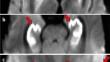
Opioid-associated brain injury may involve selective regions, including the hippocampi alone, globi pallidi, and cerebellar hemispheres. Opioid-associated amnestic syndrome, for example, is one clinical correlate of hippocampal injury as manifest by MRI abnormality. When all three regions are involved in what may be a more fulminant injury, the syndrome is termed “cerebellar, hippocampal, and basal nuclei transient edema with restricted diffusion (CHANTER)”, initially described in 2019. Until now, to our knowledge, there have been no histopathologic correlates to the imaging findings specifically in CHANTER syndrome. Here, for the first time, we present histopathologic findings of the post-mortem brain from a patient who died from complications of CHANTER syndrome following fentanyl intoxication. These observations included microhemorrhage, reactive and necrotic vasculature, eosinophilic neuronal necrosis, axonal swelling and spheroids, and frank infarction. The findings support previous experimental models implicating both hypoxic–ischemic and cytotoxic mechanisms in the tissue damage associated with CHANTER syndrome, though further work is needed to better characterize the exact cellular pathways involved to develop targeted treatments.
期刊介绍:
Acta Neuropathologica publishes top-quality papers on the pathology of neurological diseases and experimental studies on molecular and cellular mechanisms using in vitro and in vivo models, ideally validated by analysis of human tissues. The journal accepts Original Papers, Review Articles, Case Reports, and Scientific Correspondence (Letters). Manuscripts must adhere to ethical standards, including review by appropriate ethics committees for human studies and compliance with principles of laboratory animal care for animal experiments. Failure to comply may result in rejection of the manuscript, and authors are responsible for ensuring accuracy and adherence to these requirements.


 分享
分享
 求助内容:
求助内容: 应助结果提醒方式:
 扫码关注我们
