Emma Schut, Ronald M P Breedijk, Michiel F Hilbers, Mark A Hink, Tristan Krap, Maurice C G Aalders, René M Williams
{"title":"On the glow of cremated remains: long-lived green photo-luminescence of heat-treated human bones.","authors":"Emma Schut, Ronald M P Breedijk, Michiel F Hilbers, Mark A Hink, Tristan Krap, Maurice C G Aalders, René M Williams","doi":"10.1007/s43630-024-00618-2","DOIUrl":null,"url":null,"abstract":"<p><p>The long-lived green luminescence of human bone (that has been heated to 600 °C for a short duration) is attributed to a carbon quantum dot material (derived from collagen) encapsulated and protected by an inorganic matrix (derived from bone apatite) and is more intense in dense rigid and crystalline parts of (healthy) human bones. The strong collagen-apatite interaction results (upon decomposition) in a protective inorganic environment of the luminescent centers allowing long-lived triplet-based emission of a carbon (quantum) dot-like material at room temperature, as well as resilience against oxidation between 550 and 650 °C. The graphitic black phase (obtained upon heating around 400 °C) is a precursor to the luminescent carbon-based material, that is strongly interacting with the crystalline inorganic matrix. Human bone samples that have been heated to 600 °C were subjected to steady-state and time-resolved spectroscopy. Excitation-emission matrix (EEM) luminescence spectroscopy revealed a broad range of excitation and emission wavelengths, indicating a heterogeneous system with a broad density of emissive states. The effect of low temperature on the heat-treated bone was studied with Cryogenic Steady State Luminescence Spectroscopy. Cooling the bone to 80 K leads to a slight increase in total emission intensity as well as an intensity increase towards to red part of the spectrum, incompatible with a defect state model displaying luminescent charge recombination in the inorganic matrix. Time-resolved spectroscopy with an Optical Multichannel Analyzer (OMA) and Time Correlated Single Photon Counting (TCSPC) of these samples showed that the decay could be fitted with a multi-exponential decay model as well as with second-order decay kinetics. Confocal Microscopy revealed distinct (plywood type) structures in the bone and high intensity-fast decay areas as well as a spatially heterogeneous distribution of green and (fewer) red emissive species. The use of the ATTO 565 dye aided in bone-structure visualization by chemical adsorption. Conceptually our data interpretation corresponds to previous reports from the material science field on luminescent powders.</p>","PeriodicalId":98,"journal":{"name":"Photochemical & Photobiological Sciences","volume":" ","pages":"1641-1657"},"PeriodicalIF":3.2000,"publicationDate":"2024-09-01","publicationTypes":"Journal Article","fieldsOfStudy":null,"isOpenAccess":false,"openAccessPdf":"","citationCount":"0","resultStr":null,"platform":"Semanticscholar","paperid":null,"PeriodicalName":"Photochemical & Photobiological Sciences","FirstCategoryId":"92","ListUrlMain":"https://doi.org/10.1007/s43630-024-00618-2","RegionNum":3,"RegionCategory":"化学","ArticlePicture":[],"TitleCN":null,"AbstractTextCN":null,"PMCID":null,"EPubDate":"2024/9/2 0:00:00","PubModel":"Epub","JCR":"Q3","JCRName":"BIOCHEMISTRY & MOLECULAR BIOLOGY","Score":null,"Total":0}
引用次数: 0
Abstract
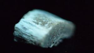
The long-lived green luminescence of human bone (that has been heated to 600 °C for a short duration) is attributed to a carbon quantum dot material (derived from collagen) encapsulated and protected by an inorganic matrix (derived from bone apatite) and is more intense in dense rigid and crystalline parts of (healthy) human bones. The strong collagen-apatite interaction results (upon decomposition) in a protective inorganic environment of the luminescent centers allowing long-lived triplet-based emission of a carbon (quantum) dot-like material at room temperature, as well as resilience against oxidation between 550 and 650 °C. The graphitic black phase (obtained upon heating around 400 °C) is a precursor to the luminescent carbon-based material, that is strongly interacting with the crystalline inorganic matrix. Human bone samples that have been heated to 600 °C were subjected to steady-state and time-resolved spectroscopy. Excitation-emission matrix (EEM) luminescence spectroscopy revealed a broad range of excitation and emission wavelengths, indicating a heterogeneous system with a broad density of emissive states. The effect of low temperature on the heat-treated bone was studied with Cryogenic Steady State Luminescence Spectroscopy. Cooling the bone to 80 K leads to a slight increase in total emission intensity as well as an intensity increase towards to red part of the spectrum, incompatible with a defect state model displaying luminescent charge recombination in the inorganic matrix. Time-resolved spectroscopy with an Optical Multichannel Analyzer (OMA) and Time Correlated Single Photon Counting (TCSPC) of these samples showed that the decay could be fitted with a multi-exponential decay model as well as with second-order decay kinetics. Confocal Microscopy revealed distinct (plywood type) structures in the bone and high intensity-fast decay areas as well as a spatially heterogeneous distribution of green and (fewer) red emissive species. The use of the ATTO 565 dye aided in bone-structure visualization by chemical adsorption. Conceptually our data interpretation corresponds to previous reports from the material science field on luminescent powders.


 分享
分享
 求助内容:
求助内容: 应助结果提醒方式:
 扫码关注我们
